
Biowissenschaften
Biowissenschaften
Hier können Sie Ihr Wissen, Ihre Forschungsfähigkeiten und Ihre praktischen Anwendungen der Mikroskopie in verschiedenen wissenschaftlichen Bereichen erweitern. Erfahren Sie, wie Sie präzise Visualisierung, Bildinterpretation und Forschungsfortschritte erzielen können. Hier finden Sie aufschlussreiche Informationen über fortgeschrittene Mikroskopie, Bildgebungsverfahren, Probenvorbereitung und Bildanalyse. Zu den behandelten Themen gehören Zellbiologie, Neurowissenschaften und Krebsforschung mit Schwerpunkt auf modernsten Anwendungen und Innovationen.

Overcoming Challenges with Microscopy when Imaging Moving Zebrafish Larvae
Zebrafish is a valuable model organism with many beneficial traits. However, imaging a full organism poses challenges as it is not stationary. Here, this case study shows how zebrafish larvae can be…

How to Automatically Obtain Fluorescent Cells of Interest in a Block-face
Block-face created by automatic trimming under fluorescence.
Mammalian cells of interest, stained with CellTrackerTM Green are visualized within the block-face using the UC Enuity equipped with the…

An Introduction to Laser Microdissection
The heterogeneity of histological and biological specimens often requires isolation of specific single cells or cell groups from surrounding tissue before molecular biology analysis can be carried…
 microdissected with a 10x objective (upper right). Inspection of the collection device (lower right).")
Molecular Biology Analysis facilitated with Laser Microdissection (LMD)
Extracting biomolecules, proteins, nucleic acids, lipids, and chromosomes, as well as extracting and manipulating cells and tissues with laser microdissection (LMD) enables insights to be gained into…
.")
Neuron Isolation in Spatial Context with Laser Microdissection (LMD)
After Alzheimer’s disease, Parkinson’s is the second most common progressive neurodegenerative disease. Before the first symptoms manifest, up to 70% of dopamine-releasing neurons in the mid-brain…
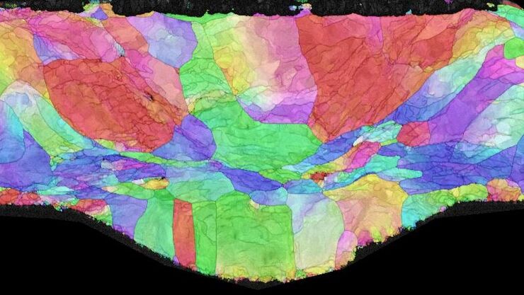
Workflow Solutions for Sample Preparation Methods for Material Science
This brochure presents and explains appropriate workflow solutions for the most frequently required sample preparation methods for material science samples.

Automatic Alignment of Sample and Knife for High Sectioning Quality
Automatic alignment of sample and knife on the ultramicrotome UC Enuity, enabling even untrained users to create ultrathin sections with reduced risk of losing precious sections.

Erforschung der Virusreplikaton mit Fluoreszenzmikroskopie
Viren können mit Hilfe verschiedener Mikroskopietechniken untersucht werden. Je nach Vergrößerung und Auflösung des Mikroskops kann die Beobachtung auf Gewebe-, Zell- oder Virionenebene erfolgen.

Epi-Illumination Fluorescence and Reflection-Contrast Microscopy
This article discusses the development of epi-illumination and reflection contrast for fluorescence microscopy concerning life-science applications. Much was done by the Ploem research group…
