Industriale
Industriale
Immergetevi in articoli dettagliati e webinar incentrati su ispezioni efficienti, flussi di lavoro ottimizzati e comfort ergonomico in contesti industriali e patologici. Gli argomenti trattati includono il controllo qualità, l'analisi dei materiali, la microscopia in patologia e molti altri. Questo è il luogo in cui potrete ottenere preziose informazioni sull'utilizzo di tecnologie all'avanguardia per migliorare la precisione e l'efficienza dei processi di produzione, nonché l'accuratezza della diagnosi e della ricerca patologica.
Fluorescent Dyes
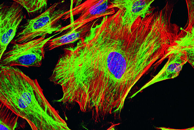
A basic principle in fluorescence microscopy is the highly specific visualization of cellular components with the help of a fluorescent agent. This can be a fluorescent protein – for example GFP –…
Fluorescence Lifetime-based Imaging Gallery
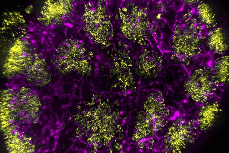
Confocal microscopy relies on the effective excitation of fluorescence probes and the efficient collection of photons emitted from the fluorescence process. One aspect of fluorescence is the emission…
Physiology Image Gallery
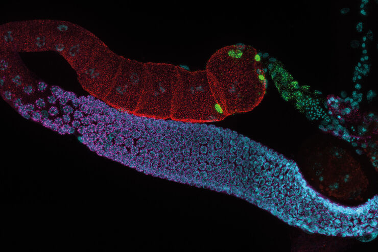
Physiology is about the processes and functions within a living organism. Research in physiology focuses on the activities and functions of an organism’s organs, tissues, or cells, including the…
Live Cell Imaging Gallery
Live cell microscopy techniques are fundamental to get a better understanding of cellular and molecular function. Today, widefield microscopy is the most common technique used to visualize cell…
Image Gallery: THUNDER Imager
To help you answer important scientific questions, THUNDER Imagers eliminate the out-of-focus blur that clouds the view of thick samples when using camera-based fluorescence microscopes. They achieve…
Organismi Modello nella Ricerca
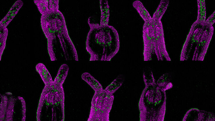
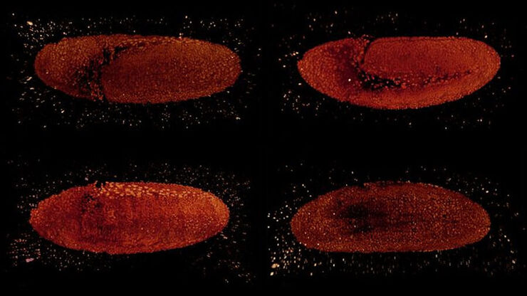
Un organismo modello è una specie utilizzata dai ricercatori per studiare specifici processi biologici. Hanno caratteristiche genetiche simili a quelle umane e vengono normalmente impiegate in aree di…
Improve 3D Cell Biology Workflow with Light Sheet Microscopy
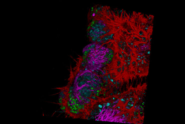
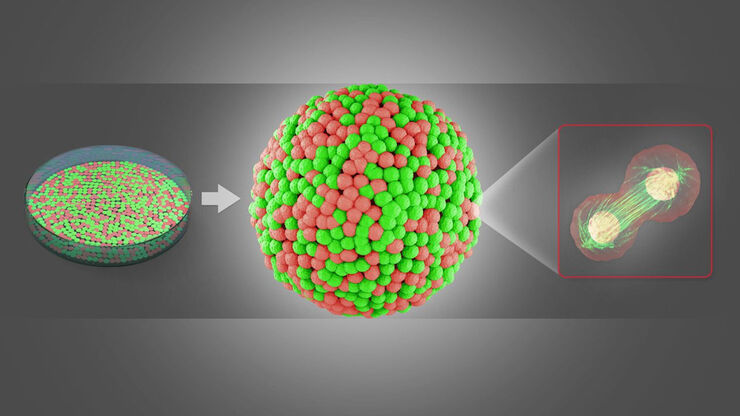
Understanding the sub-cellular mechanisms in carcinogenesis is of crucial importance for cancer treatment. Popular cellular models comprise cancer cells grown as monolayers. But this approach…
Real Time Images of 3D Specimens with Sharp Contrast Free of Haze
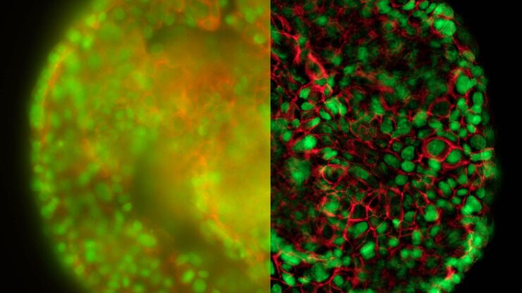
THUNDER Imagers deliver in real time images of 3D specimens with sharp contrast, free of the haze or out-of-focus blur typical of widefield systems. They can even image clearly places deep inside a…
Using a Rotation Device for Light Sheet Sample Mounting
The TCS SP8 DLS from Leica Microsystems is an innovative concept to integrate the Light Sheet Microscopy technology into the confocal microscope. Due to its unique optical architecture samples can be…
