Mica
共焦点顕微鏡
製品紹介
Home
Leica Microsystems
Mica 世界初のイメージングMicrohub。
これはすべてを変えます。
最新の記事を読む
, tubulin with Cy5 (red), and nuclei with DAPI (blue). Image courtesy of Dr. Günter Giese, Max Planck Institute for Medical Research, Heidelberg, Germany.")
Overview of Fluorescent Dyes in terms of Applications and Properties
An introduction to commonly used fluorescent dyes and an overview of their characteristics are given in this article. Fluorescence microscopy is used for the study of specific cellular components with…

History, Developments and Trends of Microscopy in Cancer Research
Cancer is a global disease, with 18 million new cases diagnosed and 10 million cancer-related deaths worldwide in 2020. This burden is set to increase, with a projected increase in cases of ~55% by…

Researchers Insights: Microscopy in Cancer Research
Discover how imaging techniques are driving cancer research forward. In this issue, we present comprehensive multimodal studies using microscopy, as well as new directions in intraoperative cancer…
, Tropomyosin (cardiomyocytes, red) and GFP (primordial cardiac layer, green).")
A Guide to Fluorescence Microscopy
Fluorescence microscopy uses the ability of fluorophores, dyes, or fluorescent proteins to emit light of a specific wavelength after being excited with light of a shorter wavelength. Biomolecules can…
 and astrocytes (green) in a cortical spheroid derived from human induced pluripotent stem cells.")
Guide to Live-Cell Imaging
For a wide range of applications in various research fields of life science, live-cell imaging is an indispensable tool for visualizing cells in a state as close to in vivo, i.e. living and active, as…

Factors to Consider When Selecting a Research Microscope
An optical microscope is often one of the central devices in a life-science research lab. It can be used for various applications which shed light on many scientific questions. Thereby the…
 and tubulin (magenta), acquired using Viventis Deep. Courtesy of Akanksha Jain, Treutlein Lab ETH-DBSSE Basel (Switzerland).")
Faster & Deeper Insights into Organoid and Spheroid Models
Gain deeper, more translatable, insights into organoid and spheroid models for drug discovery and disease research by overcoming key imaging challenges. In this eBook, explore advanced microscopy…
 at 2 weeks. Image acquired using Mica.")
How to Image Axon Regeneration in Deep Muscle Tissue
This study highlights Dr. Aaron Lee’s research on mapping nerve regeneration in muscle grafts post-amputation. Limb loss often leads to reduced quality of life, not only from tissue loss but also due…
 observed with an Ivesta 3 stereo microscope during fly pushing (sorting of the flies). The scale bar length is 1 mm. Image courtesy of M. Benton, EMBL, Heidelberg, Germany.")
A Guide to Using Microscopy for Drosophila (Fruit Fly) Research
The fruit fly, typically Drosophila melanogaster, has been used as a model organism for over a century. One reason is that many disease-related genes are shared between Drosophila and humans. It is…

神経科学研究
神経変性疾患の理解向上に取り組んでいる、もしくは神経系の機能を研究をしていますか? ライカマイクロシステムズのイメージングソリューションによってブレイクスルーを起こす方法をご覧ください。

Boston and San Francisco Innovation Hubs
Boston and San Francisco Innovation Hubs are here to help you advance scientific discovery. We provide researchers access to state-of-the-art microscope technology and expert guidance. Located in the…

Mica: A Game-changer for Collaborative Research at Imperial College London
This interview highlights the transformative impact of Mica at Imperial College London. Scientists explain how Mica has been a game-changer, expanding research possibilities and facilitating…

Overcoming Challenges with Microscopy when Imaging Moving Zebrafish Larvae
Zebrafish is a valuable model organism with many beneficial traits. However, imaging a full organism poses challenges as it is not stationary. Here, this case study shows how zebrafish larvae can be…

Exploring Microbial Worlds: Spatial Interactions in 3D Food Matrices
The Micalis Institute is a joint research unit in collaboration with INRAE, AgroParisTech, and Université Paris-Saclay. Its mission is to develop innovative research in the field of food microbiology…

Advancing Uterine Regenerative Therapies with Endometrial Organoids
Prof. Kang's group investigates important factors that determine the uterine microenvironment in which embryo insertion and pregnancy are successfully maintained. They are working to develop new…

How do Cells Talk to Each Other During Neurodevelopment?
Professor Silvia Capello presents her group’s research on cellular crosstalk in neurodevelopmental disorders, using models such as cerebral organoids and assembloids.

How to Streamline Your Histology Workflows
Streamline your histology workflows. The unique Fluosync detection method embedded into Mica enables high-res RGB color imaging in one shot.

Epi-Illumination Fluorescence and Reflection-Contrast Microscopy
This article discusses the development of epi-illumination and reflection contrast for fluorescence microscopy concerning life-science applications. Much was done by the Ploem research group…
")
Introduction to Fluorescent Proteins
Overview of fluorescent proteins (FPs) from, red (RFP) to green (GFP) and blue (BFP), with a table showing their relevant spectral characteristics.

Examining Developmental Processes In Cancer Organoids
Interview: Prof. Bausch and Dr. Pastucha, Technical University of Munich, discuss using microscopy to study development of organoids, stem cells, and other relevant disease models for biomedical…

An Introduction to Fluorescence
This article gives an introduction to fluorescence and photoluminescence, which includes phosphorescence, explains the basic theory behind them, and how fluorescence is used for microscopy.

FluoSync - a Fast & Gentle Method for Unmixing Multicolor Images
In this white paper, we focus on a fast and reliable method for obtaining high-quality multiplex images in fluorescence microscopy. FluoSync combines an existing method for hybrid unmixing with…
, SPY-Actin (cyan), and SiR-Tubulin (magenta). Instant Computational Clearing (ICC) was applied.")
How to Perform Dynamic Multicolor Time-Lapse Imaging
Live-cell imaging sheds light on diverse cellular events. As many of these events have fast dynamics, the microscope imaging system must be fast enough to record every detail. One major advantage of…
 stained to show the nucleus")
3D Spatial Analysis Using Mica's AI-Enabled Microscopy Software
This video offers practical advice on the extraction of publication grade insights from microscopy images. Our special guest Luciano Lucas (Leica Microsystems) will illustrate how Mica’s AI-enabled…

3D Tissue Imaging: From Fast Overview To High Resolution With One Click
3D Tissue imaging is a widespread discipline in the life sciences. Researchers use it to reveal detailed information of tissue composition and integrity, to make conclusions from experimental…

How To Perform Fast & Stable Multicolor Live-Cell Imaging
With the help of live-cell imaging researchers gain insights into dynamic processes of living cells up to whole organisms. This includes intracellular as well as intercellular activities. Protein or…

Simplifying Complex Fluorescence Multiwell Plate Assays
Apoptosis, or programmed cell death, occurs during organism embryo development to eliminate unwanted cells and during healing in adults to rid the body of damaged cells and help prevent cancer.…

Efficient Long-term Time-lapse Microscopy
When doing time-lapse microscopy experiments with spheroids, there are certain challenges which can arise. As the experiments can last for several days, prolonged sample survival must be achieved…

How to Prepare your Specimen for Immunofluorescence Microscopy
Immunofluorescence (IF) specimen preparations can be analyzed by various fluorescence microscopy techniques, depending on the research application. IF is indispensable for researchers using a simple…

Live-Cell Imaging Techniques
The understanding of complex and/or fast cellular dynamics is an important step for exploring biological processes. Therefore, today’s life science research is increasingly focused on dynamic…

Applying AI and Machine Learning in Microscopy and Image Analysis
Prof. Emma Lundberg is a professor in cell biology proteomics at KTH Royal Institute of Technology, Sweden. She is also the director of the Cell Atlas, an integral part of the Swedish-based Human…

Using Machine Learning in Microscopy Image Analysis
Recent exciting advances in microscopy technologies have led to exponential growth in quality and quantity of image data captured in biomedical research. However, analyzing large and increasingly…
The AI-Powered Pixel Classifier
Achieving reproducible results manually requires expertise and is tedious work. But now there is a way to overcome these challenges by speeding up this analysis to extract the real value of the image…
, Astrocytes (GFAP, red), Nuclei (DAPI, blue).")
Multicolor Microscopy: The Importance of Multiplexing
The term multiplexing refers to the use of multiple fluorescent dyes to examine various elements within a sample. Multiplexing allows related components and processes to be observed in parallel,…

Considerations for Multiplex Live Cell Imaging
Simultaneous multicolor imaging for successful experiments: Live-cell imaging experiments are key to understand dynamic processes. They allow us to visually record cells in their living state, without…

A New Method for Convenient and Efficient Multicolor Imaging
The technique combining hyperspectral unmixing and phasor analysis was developed to simplify the process of getting images from a sample labeled with multiple fluorophores. This aggregate method…
 and astrocytes (green) in a cortical spheroid derived from human induced pluripotent stem cells.")
Neuroscience Images
Neuroscience commonly uses microscopy to study the nervous system’s function and understand neurodegenerative diseases.

研究におけるモデル生物
モデル生物とは、特定の生物学的プロセスを研究するために研究者が使用する生物種です。 モデル生物は、人間と似た遺伝的特徴を持ち、遺伝子学、発生生物学、神経科学などの研究分野で一般的に使用されています。 通常、モデル生物は実験環境での維持や繁殖が容易であること、生殖サイクルが短いこと、または、特定の形質や病気を研究するために突然変異体を生成する能力を持つことで選ばれます。

がん研究
がんは、成長調節における欠損細胞によって引き起こされる複雑な異質性疾患です。 細胞または細胞群内の遺伝的および後成的変化が通常の機能を妨げ、自律的、非制御の細胞成長と増殖を引き起こします。

Introduction to Widefield Microscopy
This article gives an introduction to widefield microscopy, one of the most basic and commonly used microscopy techniques. It also shows the basic differences between widefield and confocal…
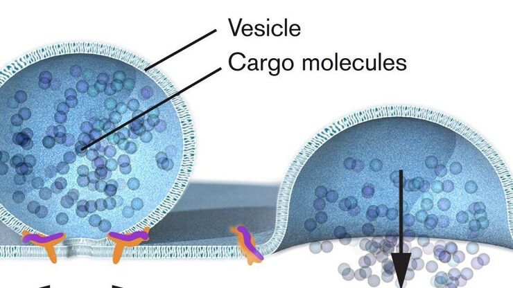
Nobel Prize 2013 in Physiology or Medicine for Discoveries of the Machinery Regulating Vesicle Traffic
On October 7th 2013, The Nobel Assembly at Karolinska Institutet has decided to award The Nobel Prize in Physiology or Medicine 2012 jointly to James E. Rothman, Randy W. Schekman and Thomas C. Südhof…

Nobel Prize 2012 in Physiology or Medicine for Stem Cell Research
The Nobel Prize recognizes two scientists who discovered that mature, specialised cells can be reprogrammed to become immature cells capable of developing into all tissues of the body. Their findings…
 inoculated with cowpea mosaic virus (CPMV) containing the GFP-gene inserted between the movement protein (MP) and the capsid proteins (CPs) in the viral RNA 2")
Introduction to Live-Cell Imaging
The understanding of complex and fast cellular dynamics is an important step to get insight into biological processes. Therefore, today’s life science research more and more demands studying…
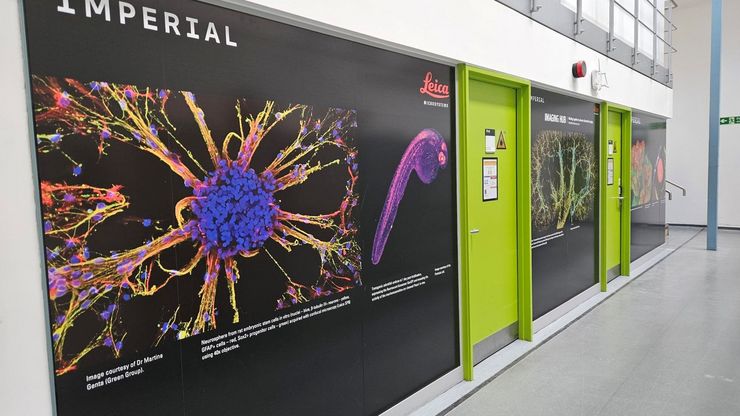
Imperial College London & Leica Microsystems Imaging Hub
Throughout its history, Leica has enthusiastically developed relationships with academic and scientific research institutions to advance scientific understanding through microscopy. Discover how you…
細胞解析
最先端の顕微鏡イメージングソリューションとAI駆動型解析ソフトウェアにより、細胞機能やサブセルラー構造の理解をより深く、より鮮明に。研究者がこれまで見えなかった生命のダイナミクスを捉え、新たな発見へと導きます。

高度な組織イメージングおよび解析
Leica Microsystems の高度なイメージングソリューションを活用することで、組織の構造および機能に対する理解を深め、空間生物学や疾患機構の解明を促進します。
バイオファーマ
ライカのソリューションは、バイオ医薬品業界において、創薬の促進、細胞解析の強化、規制を満たすデータの完全性をサポートします。
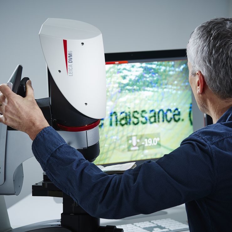
デジタル顕微鏡
デジタル顕微鏡は、接眼レンズのない顕微鏡です。 デジタルカメラが内蔵されています。 ライブ像がモニター画面に表示され、複数人で共有、画像保存、レポート出力に適しています。
Fields of Application
細胞生物学
ヒトの健康と病気を細胞ベースで理解することを目的として研究を行う場合、関心のある細胞の構造および分子の詳細から対象の細胞を研究することが重要です。 その結果、細胞生物学における顕微鏡はかってないほどに重要なツールとなり、構造環境内で試料を詳細に調査したり、細胞内小器官や高分子を分析したりすることができます。 細胞生物学イメージングは、さまざまな光電子相関顕微鏡を使用して行われます。…
細胞解析
最先端の顕微鏡イメージングソリューションとAI駆動型解析ソフトウェアにより、細胞機能やサブセルラー構造の理解をより深く、より鮮明に。研究者がこれまで見えなかった生命のダイナミクスを捉え、新たな発見へと導きます。
バイオファーマ
ライカのソリューションは、バイオ医薬品業界において、創薬の促進、細胞解析の強化、規制を満たすデータの完全性をサポートします。
高度な組織イメージングおよび解析
Leica Microsystems の高度なイメージングソリューションを活用することで、組織の構造および機能に対する理解を深め、空間生物学や疾患機構の解明を促進します。
