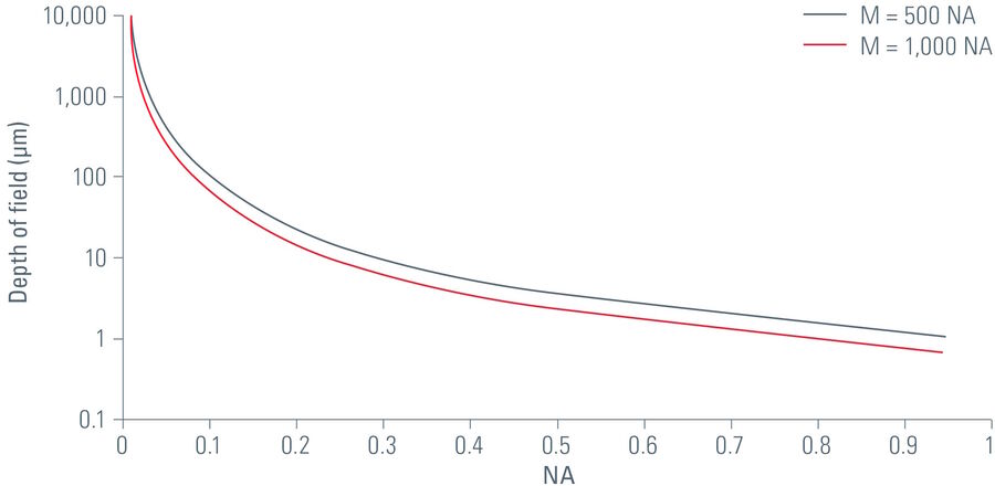
If in the above equation, the total visual magnification is replaced by the useful range of magnification with white light (λ = 550 nm) illumination, i.e., 500 x NA < MTOT VIS > 1,000 x NA [3], it can be seen that, to a first approximation, the depth of field is inversely proportional to the square of the numerical aperture.
Particularly at low magnifications, the depth of field can be significantly increased by stopping down, i.e. reducing the numerical aperture, which is normally done with the aperture diaphragm or a diaphragm on a conjugated plane. However, the smaller the numerical aperture, the lower the lateral resolution.
It is, therefore, a matter of finding the optimum balance of resolution and depth of field depending on the structure of the object. With their high-resolution objectives (high NA) and adjustable aperture diaphragms, modern light microscopes enable flexible matching of the optics to the requirements of the particular sample.
Stereo microscope observation: Depth of field and depth perception
Depth perception is greatly enhanced when observation is done via binocular rather than monocular vision, i.e., with both eyes simultaneously and not just one [4]. Disparity between the images of the same object (or group of objects) which form separately on the retina of each eye plays an important role in depth perception.
An example illustrating this phenomenal capability of the human brain is visualization of a sample with a Greenough stereo microscope. For this case, the sample-observation planes of the left and right light channels are at a slight angle to each other. In the overall image (refer to figure 2), the entire hatched area appears to be sharply focused, although this is not actually the case for either of the images seen by the left or right eye.
Stereo microscopes are often used to observe larger samples or specimens compared to compound microscopes. So it is often necessary to make a certain compromise in favor of higher depth of field, as the vertical z dimension of three-dimensional structures on such samples frequently demands it.
More depth of field and more resolution with stereo microscopy
A sophisticated approach of Leica Microsystems that gets around the inverse correlation between resolution and depth of field for stereo microscopes is FusionOptics [5]. While an observer is looking through the eyepieces, one eye sees a light channel which provides an image of the sample with higher resolution and lower depth of field. At the same time, via the second light channel, the other eye sees an image of the same sample with lower resolution and higher depth of field.
The human brain can combine the two separate images into one optimal overall image that features both higher resolution and higher depth of field.
Greater depth of field with digital imaging
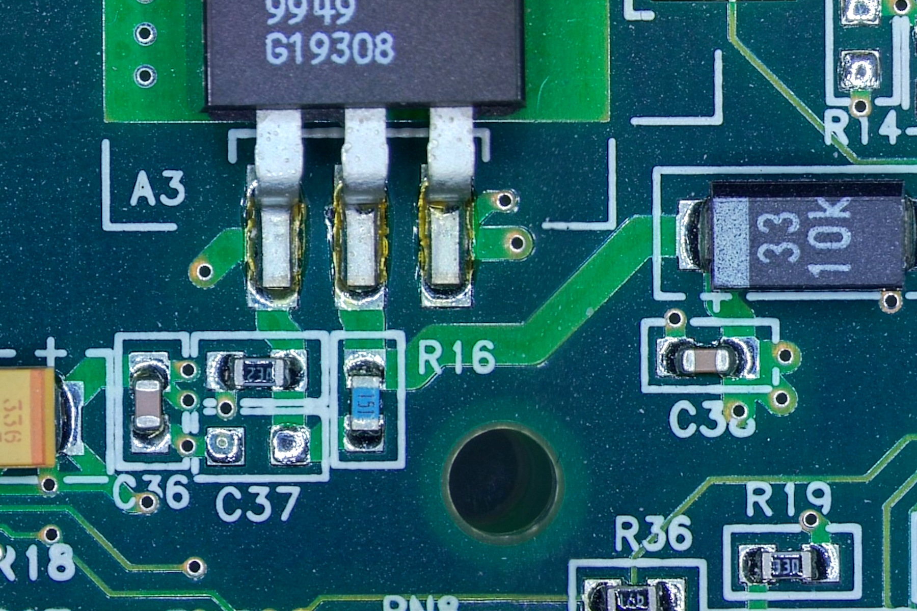
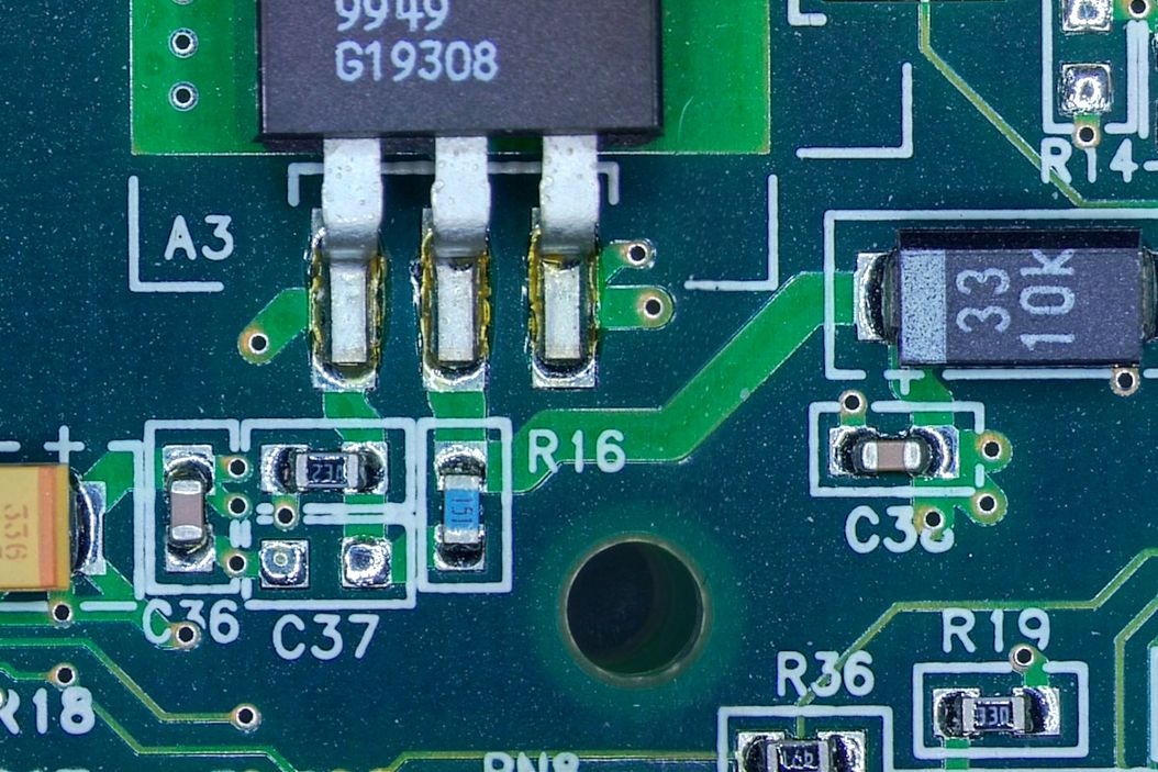
The depth of field of an optical microscope can be extended many times over with digital imaging, i.e., a digital camera is installed on the microscope for image acquistion. The illumination and other camera parameters can be set individually to optimize the quality of the resulting image.
The LAS X software provides a way to capture extended depth of field (EDOF) images. The capture of z stacks of images at different focus positions together with the intelligent image-combination algorithms enables efficient acquistion and storage of sharp images in 3 dimensions (x, y, z).
