 case.")
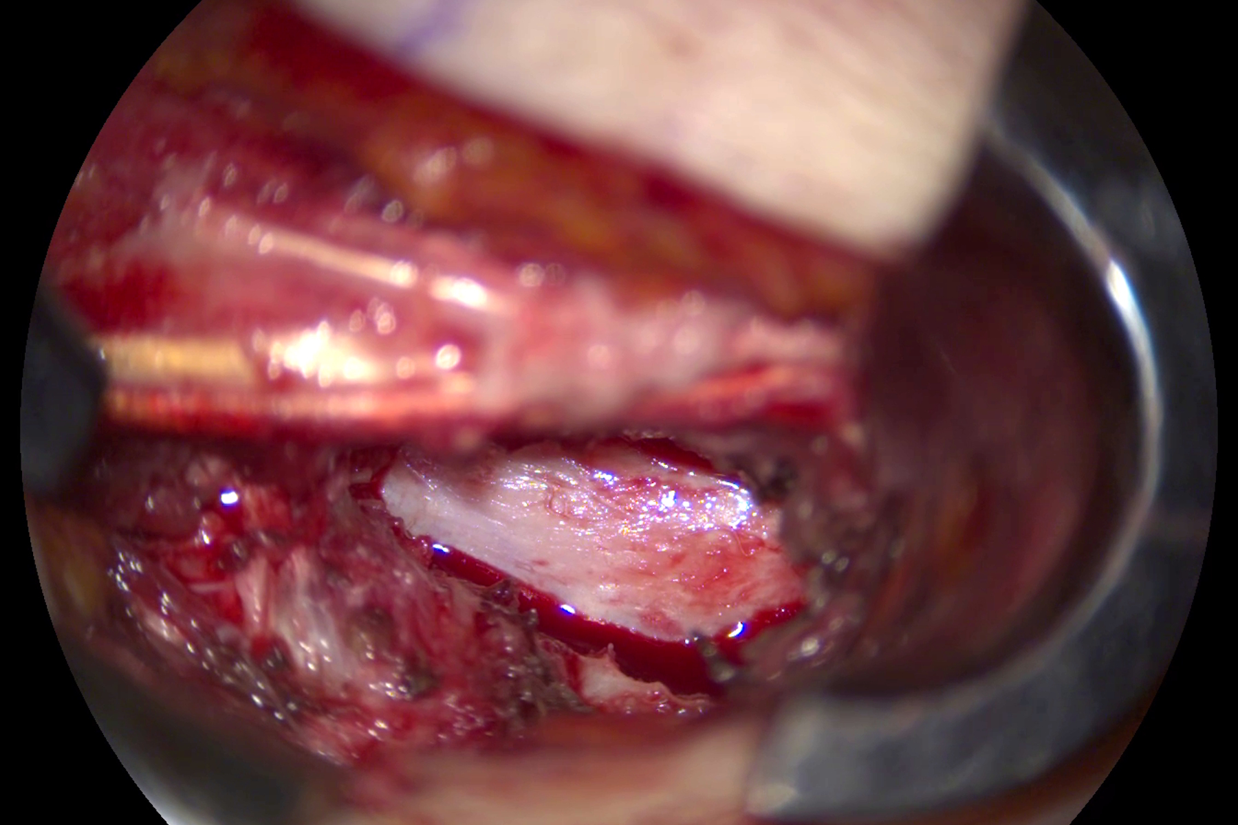
Why visualization matters in minimally invasive spine surgery
Minimal invasive spine surgery requires optimal clarity to navigate small anatomical corridors with minimal disruption to surrounding tissues [1]. Surgical microscopes help provide high magnification and superior illumination, allowing surgeons to clearly identify delicate structures such as nerves and vessels [2].
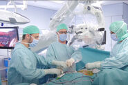
Key expert interview with Prof. Alex Alfieri, Cantonal Hospital Winterthur, Switerland
Transcript of the video
Efficiency, work comfort, and overall patient satisfaction are essential for surgeons. With the introduction of minimally invasive spine surgery, the intraoperative control of neuroanatomical structures has been improved and what is clearly evident in everyday life is that patient pain has been reduced. They can be discharged and go home within a few days. The length of stay has been reduced by 80% over the last 15 years and the outcome has improved significantly.
With the ARveo 8, my team and I have found a very stable and reliable system. The working distance, in my opinion, is one of the most important criteria for selecting a microscope. And the ARveo 8 provides a great working distance, which allows the use of all conventional instruments in the lumbar spine area, as well as in the thoracic and cervical spine area.
During the operation, we truly appreciated the opportunity to operate with a high degree of flexibility. The small angle illumination was very helpful for ensuring optimal visualization in every corner. My team and I really like the color accuracy and lighting options, always optimal even at maximum magnification. This enables us to distinguish between pathological structures and normal neuroanatomy.
Another important feature of the ARveo 8 is its excellent depth of field for the area you are working on, while also keeping the periphery in focus and that is very useful for controlling any bleeding in the area.
In spinal surgery training, it is very important not only to have good visualization, but, also, to be able to work safely in the surgical field, and the ARveo 8 makes this possible. It is particularly flexible and allows the integration of two additional eyepieces, enabling the team to work together.
You can also work heads-up with 3D-glasses looking at the big monitor, and you can change that in just a few simple steps. All handles, the foot switch, and also the mouth switch can be completely set up the way you prefer so that everything is ready for use when you enter the operating room.
Surgical microscope use in minimally invasive spine surgery
MISS requires optimal clarity to navigate small anatomical corridors with minimal disruption to surrounding tissues [3]. With the ARveo 8x, the next-generation 3D digital microscope by Leica Microsystems, surgeons can benefit from:
- Enhanced depth of field: FusionOptics technology unites an enhanced depth of field with high resolution, eliminating the need to constantly refocus and allowing surgeons to see critical structures clearly across varying depths.
- Premium optics and illumination: The combination of 400 W xenon light and Small Angle Illumination (SAI) distributes light more evenly and reduces shadows in deep and narrow cavities, ensuring a bright, fully-focused view into deeper confined surgical areas. In addition, BrightCare Plus, an integrated illumination safety function protects sensitive tissue during procedures from high light exposure. Highly light-transmissive Leica optics ensure maximum light provision, allowing safer operation at optimal light levels with clear and detail-rich image quality.
- 3D visualization: With our ARveo 8x 3D digital microscope, surgeons and the OR team can experience 3D depth perception onscreen or with the MyVeo all-in-one surgical visualization headset,
facilitating understanding of spatial information and allowing everyone to follow the surgical course more easily. The headset offers also great peripheral sight, allowing those involved in the surgery to still see their hands, the instruments, and stay connected with their team.
Conclusion
As Prof. Alfieri’s testimonial shows, the right surgical microscope is key to advancing minimally invasive spine surgery. Watch his full video to learn more about the ARveo 8’s impact on surgical practice and patient care.
Disclaimer:
The statements of the healthcare professionals included in this presentation reflect only their opinion and personal experience. Not all products are approved or offered in every country. Please contact your Leica representative for more information.