
Industrial
Industrial
Sumérjase en artículos detallados y seminarios web centrados en la inspección eficaz, los flujos de trabajo optimizados y la comodidad ergonómica en contextos industriales y patológicos. Los temas tratados incluyen el control de calidad, el análisis de materiales y la microscopía en patología, entre muchos otros. Este es el lugar donde obtendrá información valiosa sobre el uso de tecnologías de vanguardia para mejorar la precisión y la eficacia de los procesos de fabricación, así como el diagnóstico y la investigación patológicos precisos.

Polarizing Microscope Image Gallery
How polarization microscope images can be used for analysis is shown in this gallery. Polarized light microscopy (also known as polarizing microscopy) is an important method for different fields and…

Automotive Part Verification and Development according to Specifications
Automotive part verification during the development and production of parts and components by suppliers or manufacturers is important for ensuring that specifications are met. Specifications are…

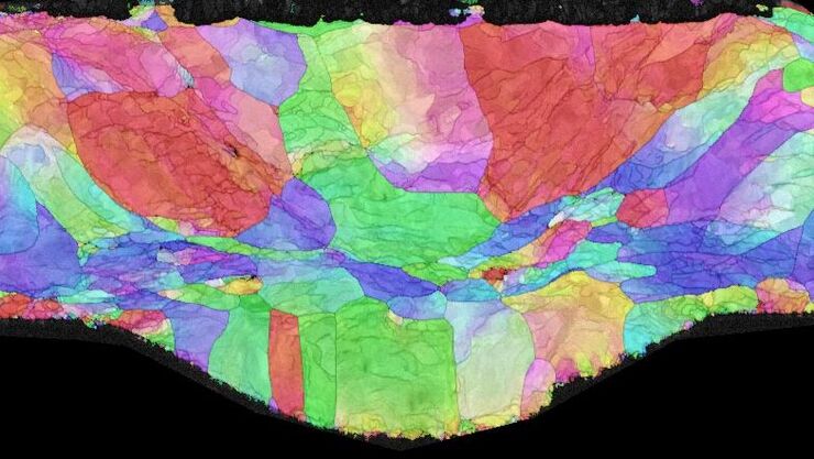
The Polarization Microscopy Principle
Polarization microscopy is routinely used in the material and earth sciences to identify materials and minerals on the basis of their characteristic refractive properties and colors. In biology,…
Workflow Solutions for Sample Preparation Methods for Material Science
This brochure presents and explains appropriate workflow solutions for the most frequently required sample preparation methods for material science samples.

Battery Particle Detection During the Production Process
How battery particle detection and analysis is enhanced with optical microscopy and laser spectroscopy for rapid, reliable, and cost-effective QC during battery production is explained in this…

Key Factors for Efficient Cleanliness Analysis
An overview of the key factors necessary for technical cleanliness and efficient cleanliness analysis concerning automotive and electronics manufacturing and production is provided in this article.

Technical Terms for Digital Microscope Cameras and Image Analysis
Learn more about the basic principles behind digital microscope camera technologies, how digital cameras work, and take advantage of a reference list of technical terms from this article.
 and oblique (right) brightfield illumination using a Leica compound microscope. The defect on the wafer surface is clearly more visible with oblique illumination.")
Rapid Semiconductor Inspection with Microscope Contrast Methods
Semiconductor inspection during the production of patterned wafers and ICs (integrated circuits) is important for identifying and minimizing defects. To increase the efficiency of quality control in…

Quality Control via Cross Sections of PCBs, PCBAs, ICs, and Batteries
Why cross sections of printed circuit boards (PCBs) and assemblies (PCBAs), integrated circuits (ICs), and battery components are useful for quality control (QC), failure analysis (FA), and research…
