
Industriale
Industriale
Immergetevi in articoli dettagliati e webinar incentrati su ispezioni efficienti, flussi di lavoro ottimizzati e comfort ergonomico in contesti industriali e patologici. Gli argomenti trattati includono il controllo qualità, l'analisi dei materiali, la microscopia in patologia e molti altri. Questo è il luogo in cui potrete ottenere preziose informazioni sull'utilizzo di tecnologie all'avanguardia per migliorare la precisione e l'efficienza dei processi di produzione, nonché l'accuratezza della diagnosi e della ricerca patologica.
.")
How Fluorescence Guides Sectioning of Resin-embedded EM Samples
Electron microscopes, including transmission electron microscopes (TEM) and scanning electron microscopes (SEM), are widely utilized to gain detailed structural information about biological samples or…

How to Save Time and Samples by Automated Ultramicrotomy
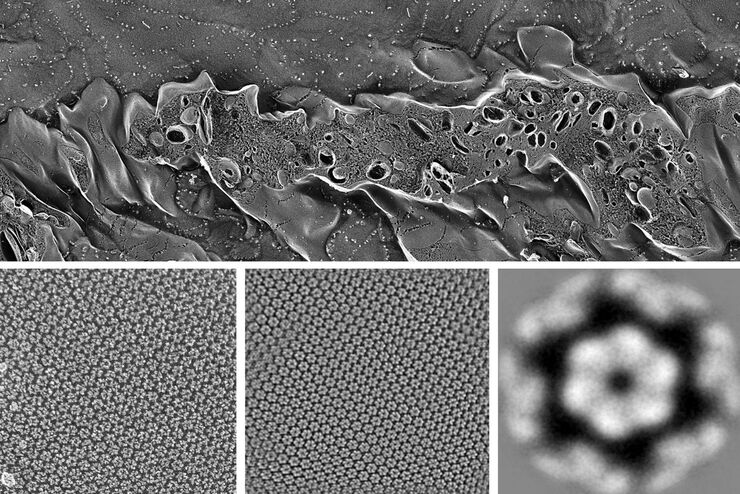
This article describes how 3D micro-CT data of a resin-embedded electron microscopy sample can be used to trim the specimen down to a defined target plane prior to sectioning. The interactive and…

How Marine Microorganism Analysis can be Improved with High-pressure Freezing
In this application example we showcase the use of EM-Sample preparation with high pressure freezing, freeze substiturion and ultramicrotomy for marine biology focusing on ultrastructural analysis of…
Advancing Cellular Ultrastructure Research
Freeze-fracture and freeze-etching are useful tools for studying flexible membrane-associated structures such as tight junctions or the enteric glycocalyx. Freeze-fracture and etching are two…

Targeting Active Recycling Nuclear Pore Complexes using Cryo Confocal Microscopy
In this article, how cryo light microscopy and, in particular cryo confocal microscopy, is used to improve the reliability of cryo EM workflows is described. The quality of the EM grids and samples is…

Essential Guide to Ultramicrotomy
When studying samples, to visualize their fine structure with nanometer scale resolution, most often electron microscopy is used. There are 2 types: scanning electron microscopy (SEM) which images the…

Soluzioni per rivestimento a polverizzazione e criodecapaggio
Dai rivestimenti realizzati in una macchina di rivestimento a polverizzazione a vuoto ridotto a temperatura ambiente a quelli realizzati a vuoto spinto fino a quelli a temperature criogeniche, le…

High Resolution Array Tomography with Automated Serial Sectioning
The optimization of high resolution, 3-dimensional (3D), sub-cellular structure analysis with array tomography using an automated serial sectioning solution, achieving a high section density on the…

Visualization of DNA Molecules
Precise low angle rotary shadowing with heavy metals (like platinum) can be used in transmission electron microscopy (TEM) to observe molecular details of objects previously absorbed on a thin, low…
