メディカル

メディカル

メディカル

脳神経外科、眼科、形成外科、耳鼻咽喉科、歯科の専門医向けに厳選された、最新の科学・臨床リソースを探索しませんか? 貴重な症例や洞察、シンポジウムなど、多彩なコンテンツをご覧いただけます。 手術用顕微鏡の最先端技術に焦点を当て、AR蛍光、3D視覚化、術中OCTイメージングといった革新的技術が、複雑な手術の精度向上や意思決定の最適化にどのように貢献するかをご紹介します。
Image: Adult rat brain. Neurons (Alexa Fluor488, green), Astrocytes (GFAP, red), Nuclei (DAPI, blue). Image courtesy of Prof. En Xu, Institute of Neurosciences and Department of Neurology of the Second Affiliated Hospital of Guangzhou Medical University, China.

Multicolor Microscopy: The Importance of Multiplexing

The term multiplexing refers to the use of multiple fluorescent dyes to examine various elements within a sample. Multiplexing allows related components and processes to be observed in parallel,…

A New Method for Convenient and Efficient Multicolor Imaging

The technique combining hyperspectral unmixing and phasor analysis was developed to simplify the process of getting images from a sample labeled with multiple fluorophores. This aggregate method…

Considerations for Multiplex Live Cell Imaging

Simultaneous multicolor imaging for successful experiments: Live-cell imaging experiments are key to understand dynamic processes. They allow us to visually record cells in their living state, without…

Designing your Research Study with Multiplexed IF Imaging

Multiplexed tissue analysis is a powerful technique that allows comparisons of cell-type locations and cell-type interactions within a single fixed tissue sample. It is common for researchers to ask…

How to Successfully Perform Live-Cell CLEM

The Leica Nano workflow provides a streamlined live-cell CLEM solution for getting insight bout structural changes of cellular components over time. Besides the technical handling described in the…

Be Confident in your Results with Cell DIVE Validated Antibodies

The Cell DIVE System includes a carefully curated list of hundreds of commercially available antibodies validated to offer optimal specificity and sensitivity in multiplexed imaging. That validation…
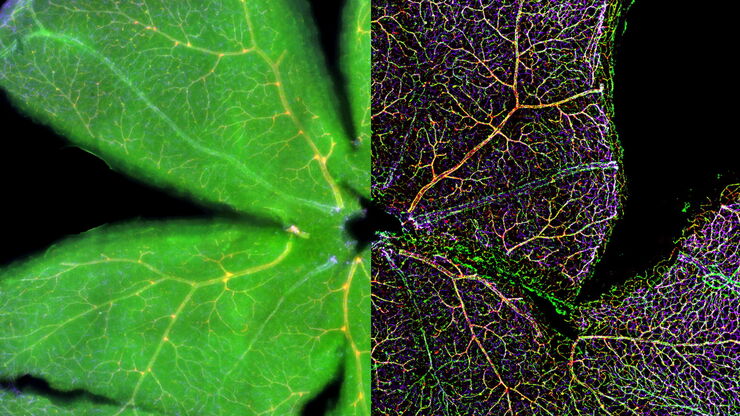
Mouse retina was fixed and stained by following reagents: anti-CD31 antibody (green): Endothelia cells, IsoB4 (red): Blood vessels, and microglia anti-GFAP antibody (blue): Astrocytes Sample courtesy by Jeremy Burton, PhD and Jiyeon Lee, PhD, Genentech Inc., South San Francisco, USA. Imaged by Olga Davydenko, PhD (Leica). Imaged with a THUNDER Imager 3D Cell Culture.

An Introduction to Computational Clearing

Many software packages include background subtraction algorithms to enhance the contrast of features in the image by reducing background noise. The most common methods used to remove background noise…
Scroll to top