Contact Us

Optical Coherence Tomography (OCT)

Leica Optical Coherence Tomography (OCT) systems support ophthalmologists, ophthalmic surgeons, and researchers with easy-to-use, high-quality imaging technology.

The preclinical OCT solutions provide insight into eye disease research, while the clinical OCT systems serve as an aid in diagnosing pathological conditions. Each system is non-invasive, contact-free, and helps detect and monitor morphological changes of ocular tissue.

OCT from Leica Microsystems

With the acquisition of Bioptigen in 2015, Leica Microsystems offers OCT imaging systems for clinical ophthalmology and preclinical research.

Need Assistance?

Contact a local specialist for expert advice on the right OCT solution for your needs and budget.

EnFocus intraoperative OCT, built into the Proveo 8 ophthalmic microscope gives surgeons greater insights, immediate confirmation, and maximum freedom in the OR. Get an immediate visual confirmation of how sub-surface tissue reacts to surgical maneuvers in crisp high-resolution OCT imaging. 

Envisu clinical OCTOCT has become a standard in the clinic, the Envisu is our noninvasive, non-contact handheld solution that aids in diagnosis and monitoring of pediatric retinal disease from neonatal infants to adults.

About Optical Coherence Tomography (OCT)

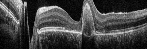
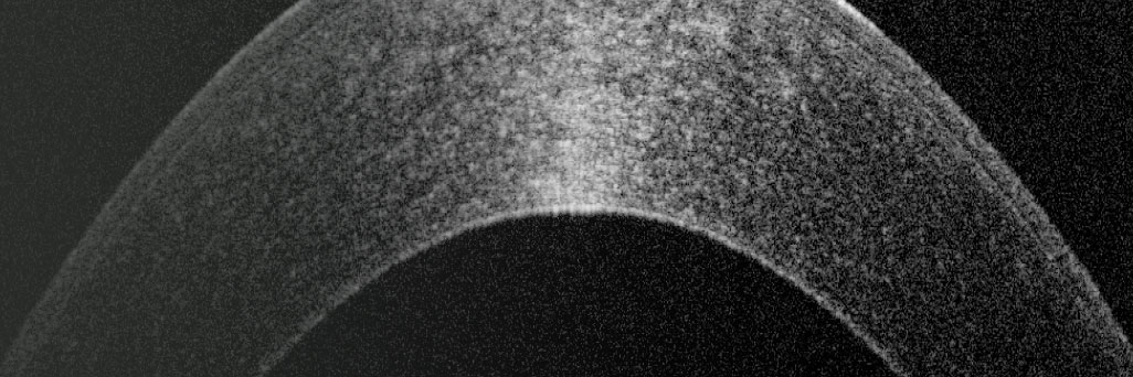
Optical coherence tomography (OCT) is an imaging technology with applications in biology, medicine, and materials investigations. In a clinical environment, OCT has revolutionized the clinical practice of ophthalmology by providing 3D anatomic information. In preclinical research, OCT facilitates early stage research into pathology and drug discovery. This non-invasive technique is used to detect and monitor morphological changes of ocular tissue, in particular retinal layer thickness, which may aid in the diagnosis of certain pathological conditions such as glaucoma, age-related macular degeneration (AMD) or diabetic retinopathy. In this Sciecne Lab topic you can find basic information about OCT, articles with application examples, case studies, and links to selected publications.

Related Articles

Read our latest articles about Optical Coherence Tomography

The knowledge portal of Leica Microsystems offers scientific research and teaching material on the subjects of microscopy. The content is designed to support beginners, experienced practitioners and scientists alike in their everyday work and experiments.

More Articles
Image of an onion flake taken with a basic Leica compound microscope after it was tested for resistance to fungus and mold growth following part 11 of the ISO 9022 standard.

ISO 9022 Standard Part 11 - Testing Microscopes with Severe Conditions

This article describes a test to determine the robustness of Leica microscopes to mold and fungus growth. The test follows the specifications of the ISO 9022 part 11 standard for optical instruments.

Immersion Objectives

How an immersion objective, which has a liquid medium between it and the specimen being observed, helps increase the numerical aperture and microscope resolution is explained in this article.
Intensity distribution (arbitrary color coding) of an image of two points where the distance between them corresponds to the Rayleigh criterion.

Microscope Resolution: Concepts, Factors and Calculation

This article explains in simple terms microscope resolution concepts, like the Airy disc, Abbe diffraction limit, Rayleigh criterion, and full width half max (FWHM). It also discusses the history.

Factors to Consider When Selecting a Research Microscope

An optical microscope is often one of the central devices in a life-science research lab. It can be used for various applications which shed light on many scientific questions. Thereby the…
Spherical aberration describes the fact that waves which pass through the centre of the lens are refracted less than the waves which pass through the edges of the curved lens.

Eyepieces, Objectives and Optical Aberrations

For most microscope applications, there are generally only two sets of optics which are adjusted by the user, namely, the objectives and the eyepieces. Of course, this is assuming that the microscope…
Steps of Koehler Illumination

Koehler Illumination: A Brief History and a Practical Set Up in Five Easy Steps

The technique of Koehler Illumination is one of the most important and fundamental techniques in achieving optimum imaging in any given light microscope set-up. Although it should be routinely used as…
Angular aperture

Collecting Light: The Importance of Numerical Aperture in Microscopy

Numerical aperture (abbreviated as ‘NA’) is an important consideration when trying to distinguish detail in a specimen viewed down the microscope. NA is a number without units and is related to the…

Optimization of the Interplay of Optical Components for Aberration Free Microscopy

Optical microscopes are used to magnify objects which are otherwise invisible for the human eye. For this purpose high quality optics is necessary to achieve appropriate resolution. However, besides…
Infinity port

Infinity Optical Systems

“Infinity Optics” refers to the concept of a beam path with parallel rays between the objective and the tube lens of a microscope. Flat optical components can be brought into this “Infinity Space”…
A 17th-century compound microscope (© Golub Collection – University of California, Berkeley/Steven Ruzin, Curator)

A Brief History of Light Microscopy

The history of microscopy begins in the Middle Ages. As far back as the 11th century, plano-convex lenses made of polished beryl were used in the Arab world as reading stones to magnify manuscripts.…
Background image
Scroll to top