Science Lab

Science Lab

Science Lab

Bem-vindo ao portal de conhecimento da Leica Microsystems. Você encontrará pesquisas científicas e material didático sobre o tema microscopia. O portal oferece suporte a iniciantes, profissionais experientes e cientistas em seus trabalhos e experimentos diários. Explore tutoriais interativos e notas de aplicação, descubra os fundamentos da microscopia, bem como as tecnologias de ponta. Faça parte da comunidade do Science Lab e compartilhe sua experiência.
TIRF Image of Tubulin, YFP, penetration depth: 120 mm

Applications of TIRF Microscopy in Life Science Research

The special feature of TIRF microscopy is the employment of an evanescent field for fluorophore excitation. Unlike standard widefield fluorescence illumination procedures with arc lamps, LEDs or…
Snapshot from a time lapse of a calcium imaging experiment using the ratiometric calcium indicator Fura-2.

Ratiometric Imaging

Many fundamental functions of a cell strongly depend on delicate, but nevertheless dynamic balances of ions (e.g. calcium, magnesium), voltage potentials and pH between the cell’s cytosol and the…

Fluorescence Recovery after Photobleaching (FRAP) and its Offspring

FRAP (Fluorescence recovery after photobleaching) can be used to study cellular protein dynamics: For visualization the protein of interest is fused to a fluorescent protein or a fluorescent dye. A…

Förster Resonance Energy Transfer (FRET)

The Förster Resonance Energy Transfer (FRET) phenomenon offers techniques that allow studies of interactions in dimensions below the optical resolution limit. FRET describes the transfer of the energy…

An Introduction to CARS Microscopy

CARS overcomes the drawbacks of conventional staining methods by the intrinsic characteristics of the method. CARS does not require labeling because it is highly specific to molecular compounds which…
Scheme of a 2D mosaic scan. Drosophila melanogaster (eye section)

Mosaic Images

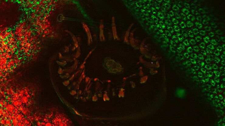
Confocal laser scanning microscopes are widely used to create highly resolved 3D images of cells, subcellular structures and even single molecules. Still, an increasing number of scientists are…

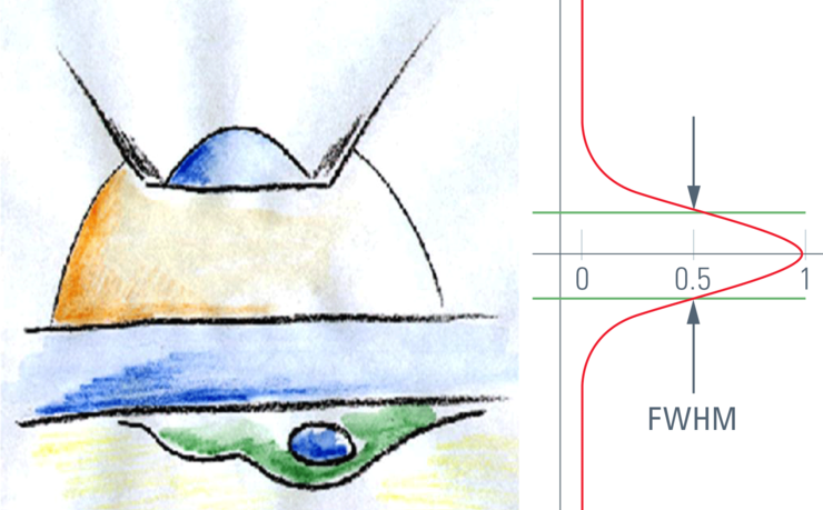
Confocal Optical Section Thickness

Confocal microscopes are employed to optically slice comparably thick samples.
Section taste buds rabbit, differential interference contrast microscope

Optical Contrast Methods

Optical contrast methods give the potential to easily examine living and colorless specimens. Different microscopic techniques aim to change phase shifts caused by the interaction of light with the…
Modulation contrast visualizes transparent, low-contrast specimens.

Integrated Modulation Contrast (IMC)

Hoffman modulation contrast has established itself as a standard for the observation of unstained, low-contrast biological specimens. The integration of the modulator in the beam path of themodern…
Scroll to top