Getting Sharper 3D Images of Thick Biological Specimens with Widefield Microscopy
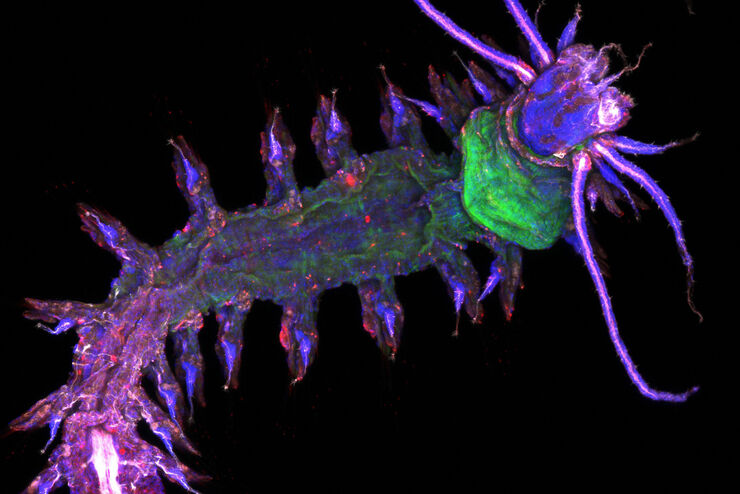
Widefield fluorescence microscopy is often used to visualize structures in life science specimens and obtain useful information. With the use of fluorescent proteins or dyes, discrete specimen…Read articleDrosophila Testis Niche Stem Cells – Three Color Computational Clearing
Differentiated living beings such as humans, but also a fruit fly or a plant, possess not only the differentiated cells which form specific tissues, but also those cells whose fate is not yet (or only…Read articleWhere to go? Cellular Migration requires coordinated Transitions of Actin Cortex
Plants, Bacteria, and Fungi possess a rigid cell wall that protects the cell and gives it shape. Animal cells, such as mammalian cells, have no outer wall, which exposes their plasma membrane to the…Read articleAlzheimer Plaques: fast Visualization in Thick Sections
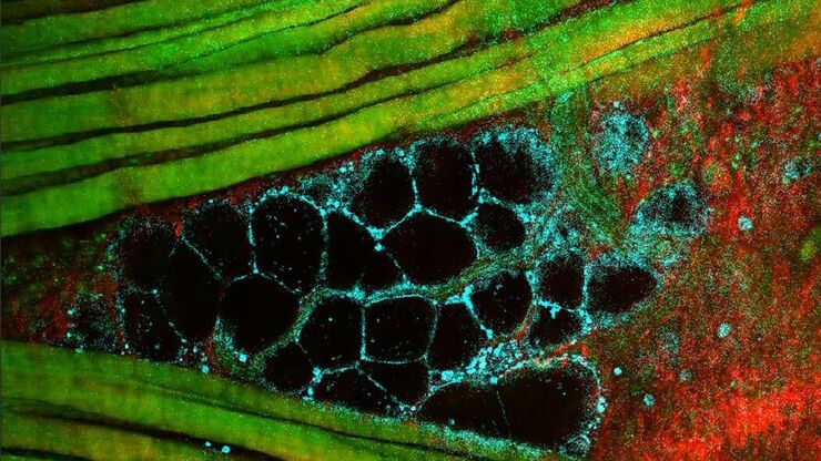
More than 60% of all diagnosed cases of dementia are attributed to Alzheimer’s disease. Typical of this disease are histological alterations in the brain tissue. So far, there is no cure for this…Read articleVisualize developmental patterns in Drosophila
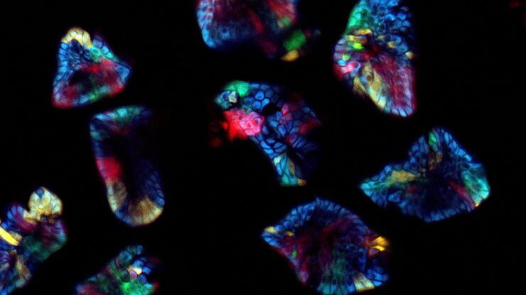
The marvelous complexity of life is perhaps the greatest source of fascination within the biological sciences. Among the most complex subjects within biology is the development of multicellular life.…Read articleZebrafish Brain - Whole Organ Imaging at High Resolution
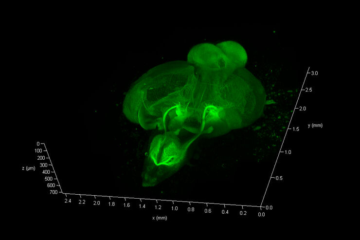
Structural information is key when one seeks to understand complex biological systems, and one of the most complex biological structures is the vertebrate central nervous system. To image a complete…Read articleWhat is a Spectral Detector (SP Detector)?
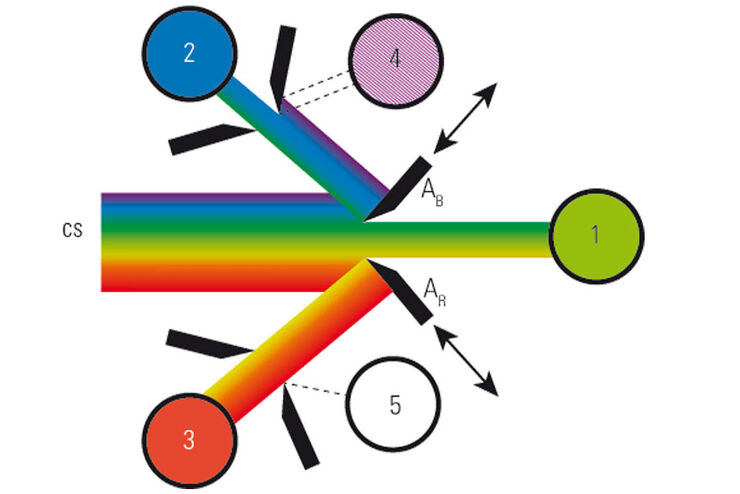
The SP detector from Leica Microsystems denotes a compound detection unit for point scanning microscopes, in particular confocal microscopes. The SP detector splits light into up to 5 spectral bands.…Read articleWhat is a Resonant Scanner?
A resonant scanner is a type of galvanometric mirror scanner that allows fast image acquisition with single-point scanning microscopes (true confocal and multiphoton laser scanning). High acquisition…Read articleWhat is a Field-of-View Scanner?
A field-of-view scanner is an assembly of galvanometric scanning mirrors used in single-point confocal microscopes that offer the correct optical recording of large field sizes. The field-of-view…Read articleResolved Field Number (RFN)
The field number (FN) for optical microscopes indicates the field of view (FOV). It corresponds to the area in the intermediate image that is observable through the eyepieces. Although, we cannot…Read articleWhat is a Tandem Scanner?
A Tandem Scanner is an assembly of two different types of scanning together in one system for true confocal point scanning. The Tandem Scanner consists of a three-mirror scanning base with the…Read articleWhich Sensor is the Best for Confocal Imaging?
The Hybrid Photodetectors (HyD) are! Why that is the case is explained in this short Science Lab article.Read articleFLIM FRET and Biosensors: Versatile Tools for Biomedical Research
Fluorescence Lifetime Imaging (FLIM) in combination with Förster Resonance Energy Transfer (FRET) has proven to be very beneficial for investigations in biomedical research for a wide range of…Read articleLifetime – a Proper Alternative
„Way too complicated!“ - the notorious feedback when it comes to fluorescence lifetime measurements. This will change now! New technologies and new concepts for data evaluation, all implemented in…Read articleMultiphoton Microscopy – a Satisfied Wish List
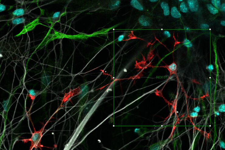
The colorful picture shows colon tumor cells, fluorescently labelled and lineage traced from a multicolor tracer. The gray color codes for the second harmonic generation (SHG) signal from Collagen 1.…Read articleMission Impossible Accomplished: Tunable Colors for Non-descanning Detection
Leica Microsystems’ 4Tune detector, the key component of the SP8 DIVE Deep In Vivo Explorer, provides spectrally tunable image recording with non-descanning detection. An innovative solution for…Read articleLaser Beam Shaping for Multicolor Multiphoton Microscopy
Multiphoton Microscopy is one of the current hot topics in life science research. The new Leica TCS SP8 DIVE from Leica Microsystems presents a series of beneficial new innovations, including a freely…Read articlePrimary Beam Splitting Devices for Confocal Microscopes
Current fluorescence microscopy employs incident illumination which requires separation of illumination and emission light. The classical device performing this separation is a color-dependent beam…Read article