Introduction
Malaria parasite development in the mosquito midgut is difficult to analyze because parasites are not uniformly distributed within host tissue.
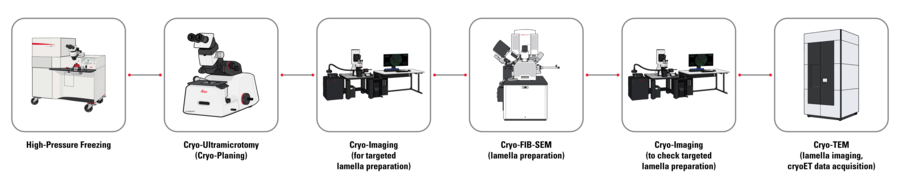
This correlative workflow combines high-pressure freezing, cryo-planing, cryo-fluorescence imaging, and cryo-EM to identify parasite-containing oocysts before lamella preparation and enable targeted cryo-electron tomography.
Identification of parasite-rich regions before lamella preparation
Mosquito midguts were dissected and transferred into high-pressure freezing planchettes prepared with yeast paste as cryoprotectant. The loaded planchettes were then high-pressure frozen with EM ICE.
DAPI staining before high-pressure freezing was used to visualize nuclei throughout the vitrified tissue and support orientation within the sample.
Fluorescence-guided cryo electron tomography (cryoET) workflow
Video: Cryo-fluorescence overview of a frozen sample carrier containing mosquito midgut tissue stained with DAPI. Regions of increased fluorescence intensity highlight areas with higher cellular density, enabling identification of potential malaria parasite-rich regions prior to cryo-planing and downstream cryo-EM analysis Sample courtesy: Nedal Darif, Postdoctoral Fellow, EMBL Heidelberg, Germany.
Cryo-planing improves the clarity of fluorescent signal in vitrified samples
Under cryogenic conditions, brightfield imaging alone provides limited information for locating internal regions of interest. Cryo-planing and trimming with the UC Enuity ultramicrotome enabled controlled removal of surface material, produced a clean block face, and progressively exposed the target region.
The workflow with the UC Enuity cryo chamber comprised initial block-face visualization by brightfield imaging, iterative cryo-planing with fluorescence inspection, and trimming to expose internal structures. After planing, the fluorescence signal was clearer, supporting more accurate localization of the region of interest.

Fluorescence imaging guides serial lamella preparation with the SOLIST method
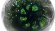
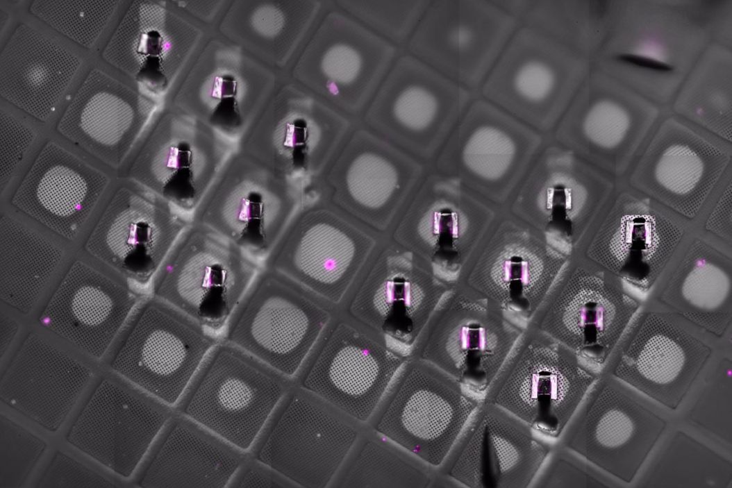
Following planing, cryo-confocal imaging with STELLARIS Cryo was used to visualize parasites within individual oocysts. These oocysts contain developing malaria parasites and define regions of interest for targeted lamella preparation.
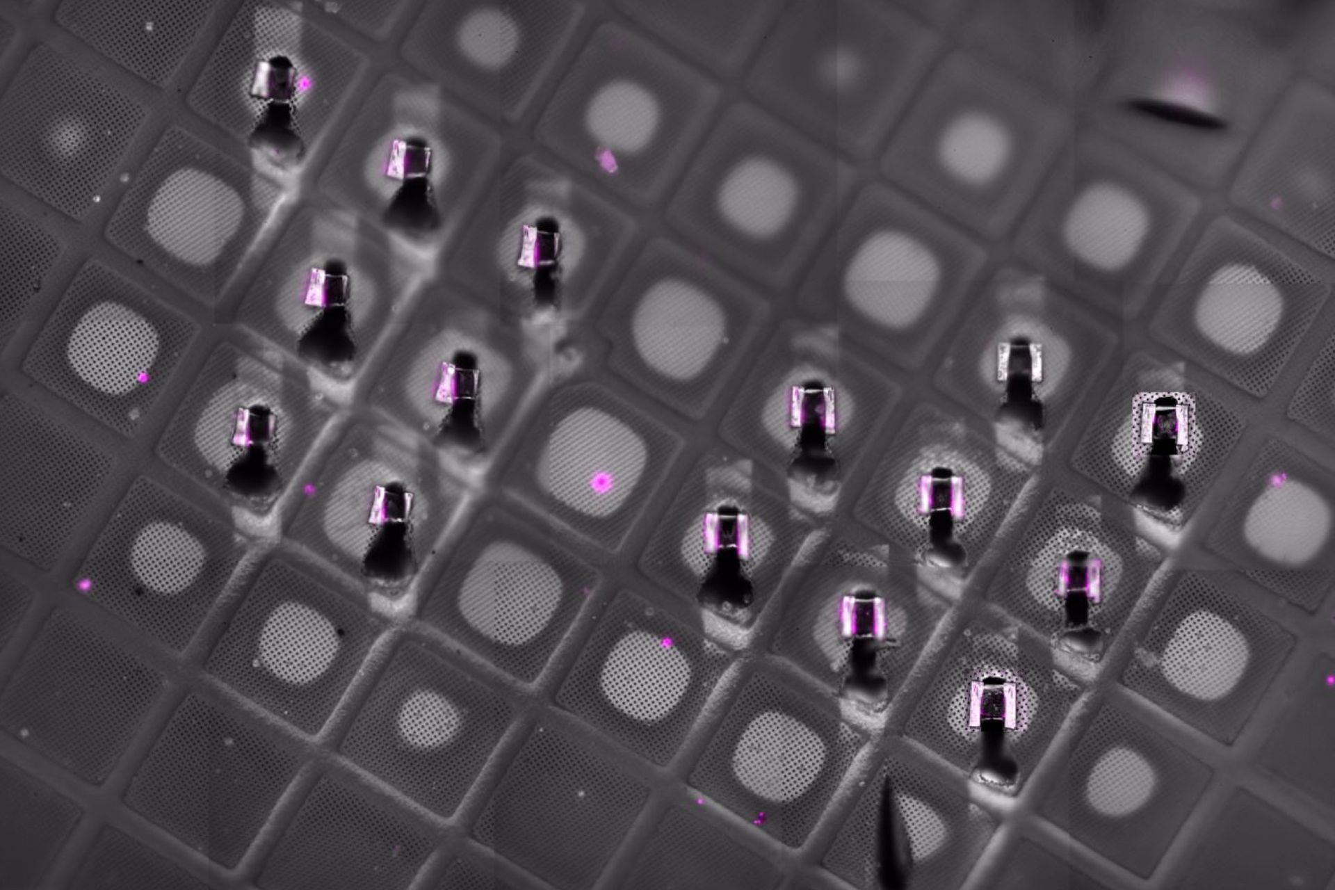
Lamellae were prepared from the identified regions on an Aquilos 2 cryo-FIB-SEM using the SOLIST method (Serialized on-grid lift-in sectioning for tomography). The lamellae were transferred onto support grids and thinned to ~200 nm by FIB milling.
Cryo-confocal imaging reveals malaria parasite-containing oocysts in midgut tissue

Targeted cryo-FIB lamella preparation from fluorescence-defined regions

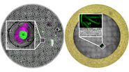
Cryo-confocal imaging of milled lamella on TEM grid confirms successful targeting

Cryo-ET reveals ultrastructural features of malaria parasites
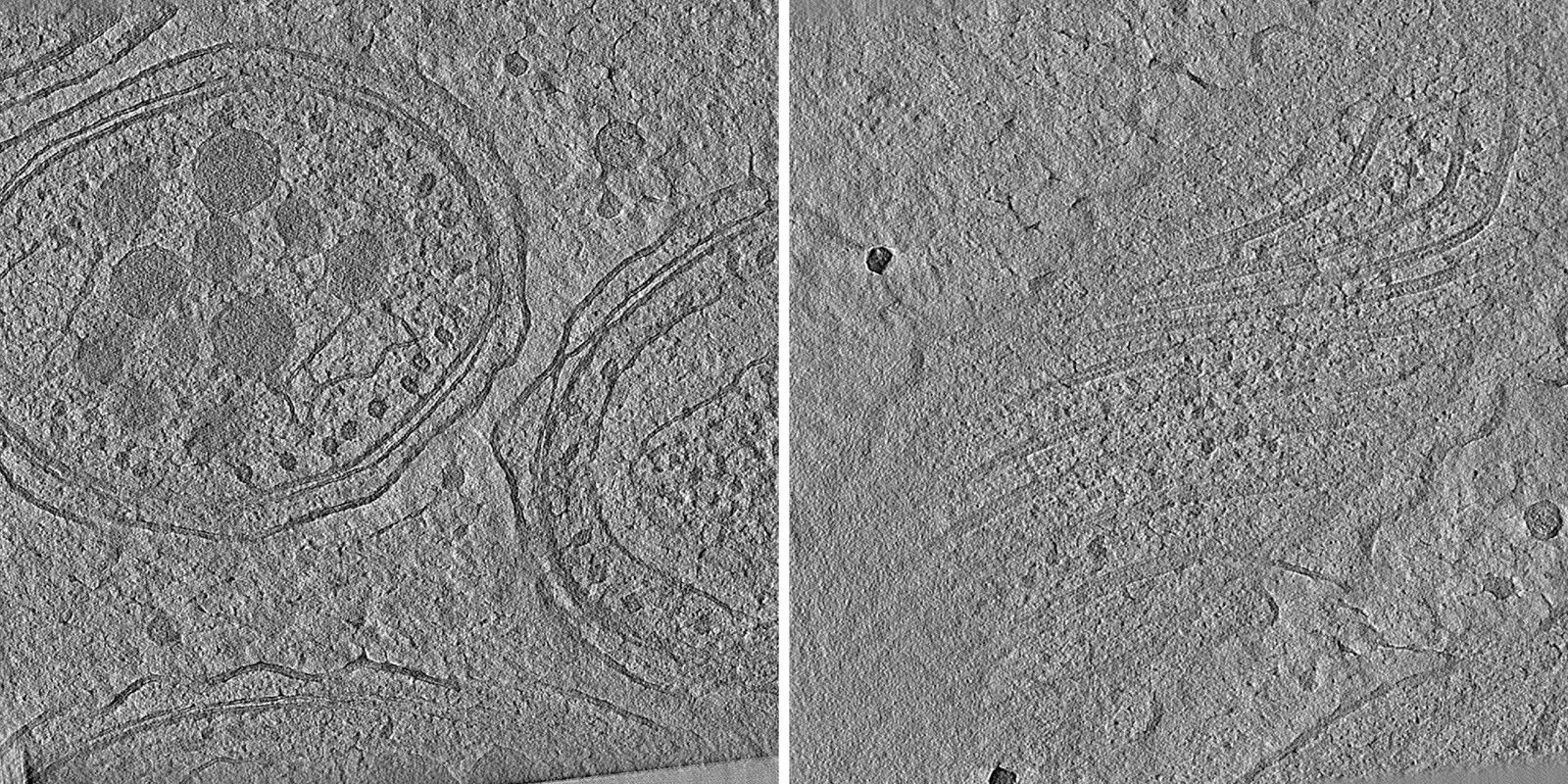
The prepared lamellae were analyzed by cryo-TEM on a Titan Krios. Cryo-ET revealed parasite morphology, membrane organization, internal structural features, and the surrounding host-tissue context.
Cryo-electron tomography of malaria parasite ultrastructure
Application value
- Enables efficient targeting to regions of interest for cryo-ET in complex biological samples
- Reduces time spent on lamella preparation and cryo-ET in non-relevant regions
- Supports integration of cryo-fluorescence and cryo-EM workflows
- Facilitates analysis of rare biological events