Surgical microscope use in minimally invasive spine surgery
MISS equires optimal clarity to navigate small anatomical corridors with minimal disruption to surrounding tissues [2]. The PROvido surgical microscope supports these procedures by offering:
- Enhanced depth of field: FusionOptics technology unites an enhanced depth of field with high resolution, eliminating the need to constantly refocus and allowing surgeons to see critical structures clearly across varying depths.
- Illumination: The combination of 300 W xenon light and Small Angle Illumination (SAI) distributes light more evenly and reduces shadows deep into narrow channels, ensuring a bright, fully-focused view deeper into confined surgical areas.
- Ergonomics: The electromagnetic brakes and AC/BC balancing allows surgeons to effortlessly position the optics carrier in the required angle. The robust, full-metal stand ensures fast stabilization and remains exactly where needed.
By providing a bright, fully-focused view deeper into narrow cavities with FusionOptics and concentrated xenon light, PROvido helps advance the efficiency of minimally invasive spine surgery.
Expert interview with Dr. Sven Bamps on surgical visualization
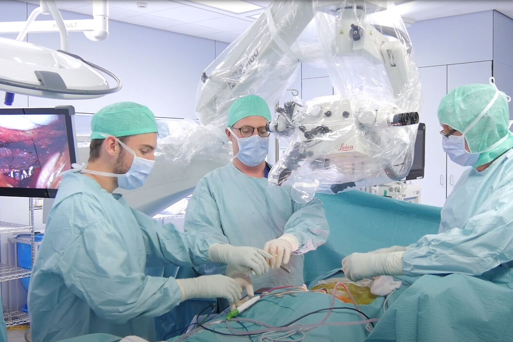
Dr. Sven Bamps, neurosurgeon at Saint-Truiden Hospital and Hasselt University (Belgium), uses the PROvido surgical microscope during spinal procedures. In a posterior lumbar fusion of the L4–L5 vertebrae, he highlighted the benefits of enhanced visualization, intuitive handling, and ergonomic design:
The microscope provides a very clear view of anatomical layers like the ligamentum flavum, nerve roots, and the dural sac. With optimized focus and zoom, I can differentiate structures with confidence, while the illumination reaches deep into the operative field.
Key learnings and benefits of the PROvido surgical microscope in spine surgery
- Clear anatomical visualization – Surgeons can distinguish ligaments, nerve roots, and the dural sac with precision.
- FusionOptics technology – A deeper field of view with high resolution reduces the need for constant refocusing, saving time during delicate spinal procedures.
- Consistent, deep illumination – SAI further improves visualization by distributing light more evenly and reducing shadows deep into narrow channels.
- Ergonomic flexibility – The floor stand and intuitive controls allow repositioning without disrupting posture, lowering fatigue in long operations.
- Collaborative visualization – The high-resolution side screen enables the OR team to follow the procedure in real time, enhancing workflow efficiency.
Video transcript: Advanced visualization in posterior lumbar fusion surgery
What kind of spine procedure did you perform with the PROvido?
“The procedure we performed was a posterior lumbar fusion of the L4 and L5 vertebrae, with a decompression of the L4 and L5 nerve root on the asymptomatic left side.”
What was your impression using the PROvido in spine surgery?
“My first impression was very good, as it’s a very nice device, easy to use, easy to handle. And the design of the machine is also very easy to use for my operation staff in the OR. For example, the touch screen is very handy, very easy to use, and we can also easily connect it to our upper screens into the operating theater.”
Which advantages did you experience during surgery using the PROvido?
“The advantages using the microscope was that I could gain a clear view of the exiting nerve roots on the damaged side, and we could have a nice vision, a very in-depth focus at the operating side. I can really see a very nice difference in the anatomical layers. I can see the yellow ligament, I can see the nerve root and the dural sac, the thecal sac, and I can clearly differentiate them while playing with the focus and the zoom.”
What did you like most about the PROvido?
“The main benefits for me is that I can bring light, nicely, deeply into the operative field, that I can change the position of the microscope without changing my physiological position so it’s easier to operate on the patient, and it’s better for the patient because we can make the procedure less invasive.”
Would you recommend the PROvido to your colleagues?
“Yes, I will suggest it to them. It’s also very user friendly into the operating theater and the side screen, the new flat screen, has a very high image quality so that everybody in the room can enjoy the operation.”
The statements of the healthcare professionals included in this presentation reflect only their opinion and personal experience. Not all products are approved or offered in every country. Please contact your Leica representative for more information.
References
- American Academy of Orthopaedic Surgeons. Minimally invasive spine surgery [Internet]. Rosemont (IL): AAOS; [updated 2024; cited 2025 Oct 2].
- American Association of Neurological Surgeons. Minimally invasive spine surgery [Internet]. Rolling Meadows (IL): AANS; [updated 2024 Apr 8; cited 2025 Oct 2].
Related Articles
-
 case.")
Flexibility and Efficiency in Minimally Invasive Spine Surgery
According to Prof. Alex Alfieri, Chief Physician and Head of clinic for Neurosurgery and Spinal…
Jan 26, 2026Read article -

Use of AR Fluorescence in Neurovascular Surgery
Learn about the use of GLOW800 Augmented Reality in neurovascular surgery through clinical cases and…
Aug 02, 2023Read article -

Neurosurgical Treatment of Spinal Arterio-Venous Fistulas
Learn about the neurosurgical treatment of spinal arterio-venous fistulas, including classification,…
Nov 23, 2022Read article
