Corporate Communications

Leica Microsystems entwickelt und fertigt Mikroskope und wissenschaftliche Instrumente für die Analyse von Mikro- und Nanostrukturen.
Wir bieten wissenschaftliches Forschungs- und Lehrmaterial zu den Themen der Mikroskopie an. Die Inhalte sind so gestaltet, dass sie Anfänger, erfahrene Praktiker und Wissenschaftler gleichermaßen bei ihrer täglichen Arbeit und ihren Experimenten unterstützen. Erkunden Sie interaktive Tutorials und Anwendungshinweise, entdecken Sie die Grundlagen der Mikroskopie ebenso wie High-End-Technologien.
Folgen Sie uns!
Advancing Cell Biology with Cryo-Correlative Microscopy
Correlative light and electron microscopy (CLEM) advances biological discoveries by merging different microscopes and imaging modalities to study systems in 4D. Combining fluorescence microscopy with…

Image Gallery: THUNDER Imager
To help you answer important scientific questions, THUNDER Imagers eliminate the out-of-focus blur that clouds the view of thick samples when using camera-based fluorescence microscopes. They achieve…

Hand-Held OCT Clinical & Research Applications
Hand-held Optical Coherence Tomography (OCT) has revolutionized pediatric ophthalmology and has had a significant impact on ophthalmology in general. The Leica Envisu C2300 hand-held OCT allows to…
From Organs to Tissues to Cells: Analyzing 3D Specimens with Widefield Microscopy
Obtaining high-quality data and images from thick 3D samples is challenging using traditional widefield microscopy because of the contribution of out-of-focus light. In this webinar, Falco Krüger…

Plastische und rekonstruktive Chirurgie: Warum ein Mikroskop benutzen?
Plastic and Reconstructive Surgery procedures can be delicate. Visualization solutions play an essential role, allowing to perform the surgery in the best conditions. And more and more plastic…

Minimally Invasive Spine Surgery: Improving Precision and Accuracy with Microscopes
Spine surgery is extremely delicate and requires extensive training and experience. Innovative visualization technologies can also help achieve better outcomes allowing to see more and have a clearer…

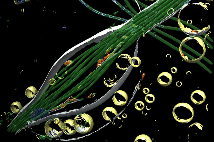
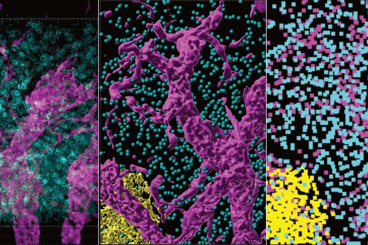
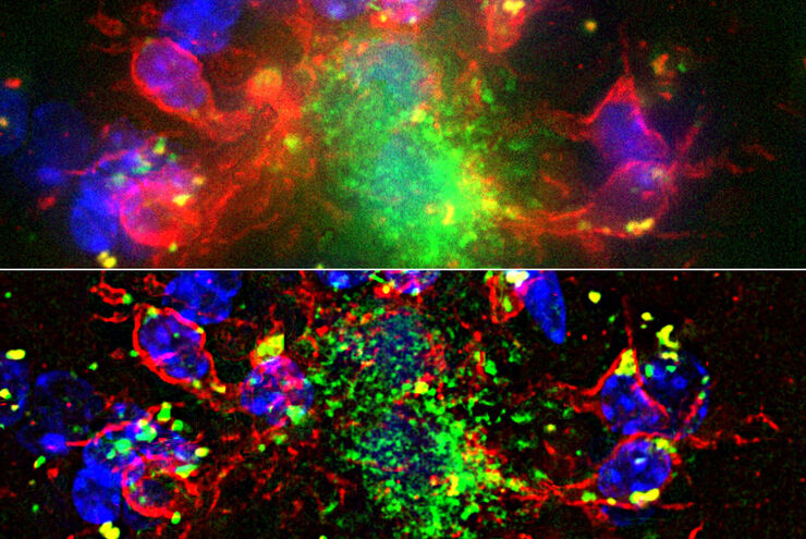
Mit LIGHTNING das Maximum an Informationen aus Ihrer Probe erhalten
LIGHTNING ist ein adaptiver Prozess zur Extraktion von Bildinformationen, bei dem vollautomatisch anderweitig nicht sichtbare Strukturen und feine Details sichtbar gemacht werden. Im Gegensatz zu…
Advanced Techniques in Cataract and Refractive Surgery
In this webinar Dr. Thompson and Dr. Moshirfar will explain how Leica microscopes aid in procedures such as Centration of Multifocal IOLs and corneal inlays such as Kamra and Lenticular Grafts used in…
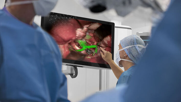
Clinical Uses in Cerebrovascular and Skull Base Neurosurgery
In this webinar Dr. Bendok and Dr. Morcos explain how Augmented Reality and Fluorescence can enhance visualization and support surgical decision making. They present first-hand experience of the GLOW…

Surgical Microscopes: Key Factors for OR Nurses
Operating room (OR) nurses are vital to the surgery process. An OR Nurse Manager explains the key surgical microscope features to facilitate their work.
Introduction to Ion Beam Etching with the EM TIC 3X
In this article you can learn how to optimize the preparation quality of your samples by using the ion beam etching method with the EM TIC 3X ion beam milling machine. A short introduction of the…
Computational Clearing - Enhance 3D Specimen Imaging
This webinar is designed to clarify crucial specifications that contribute to THUNDER Imagers' transformative visualization of 3D samples and improvements within a researcher's imaging-related…

STELLARIS White Light Lasers
When it comes to choosing fluorescent probes for your multi-color experiments, you shouldn’t have to compromise. Now you can advance beyond conventional excitation sources that limit your fluorophore…

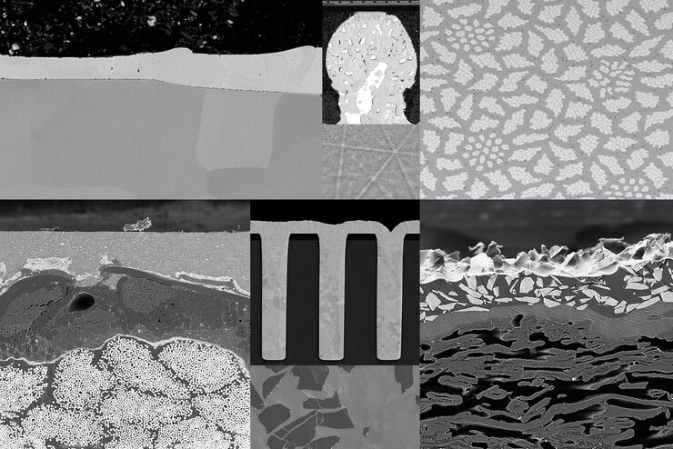
Workflow Solutions for Sample Preparation Methods for Material Science
This brochure presents and explains appropriate workflow solutions for the most frequently required sample preparation methods for material science samples.

How to Drape an Overhead Surgical Microscope
The tutorial features the Leica ARveo digital Augmented Reality microscope for complex neurosurgery. The procedure also applies to the Leica M530 OHX, OH6, OH5 and OH4.

How to Drape a Surgical Microscope
Before performing surgical procedures, it is important to drape the surgical microscope to ensure sterile working conditions. At Leica, we are committed to helping you with your surgical practice. In…
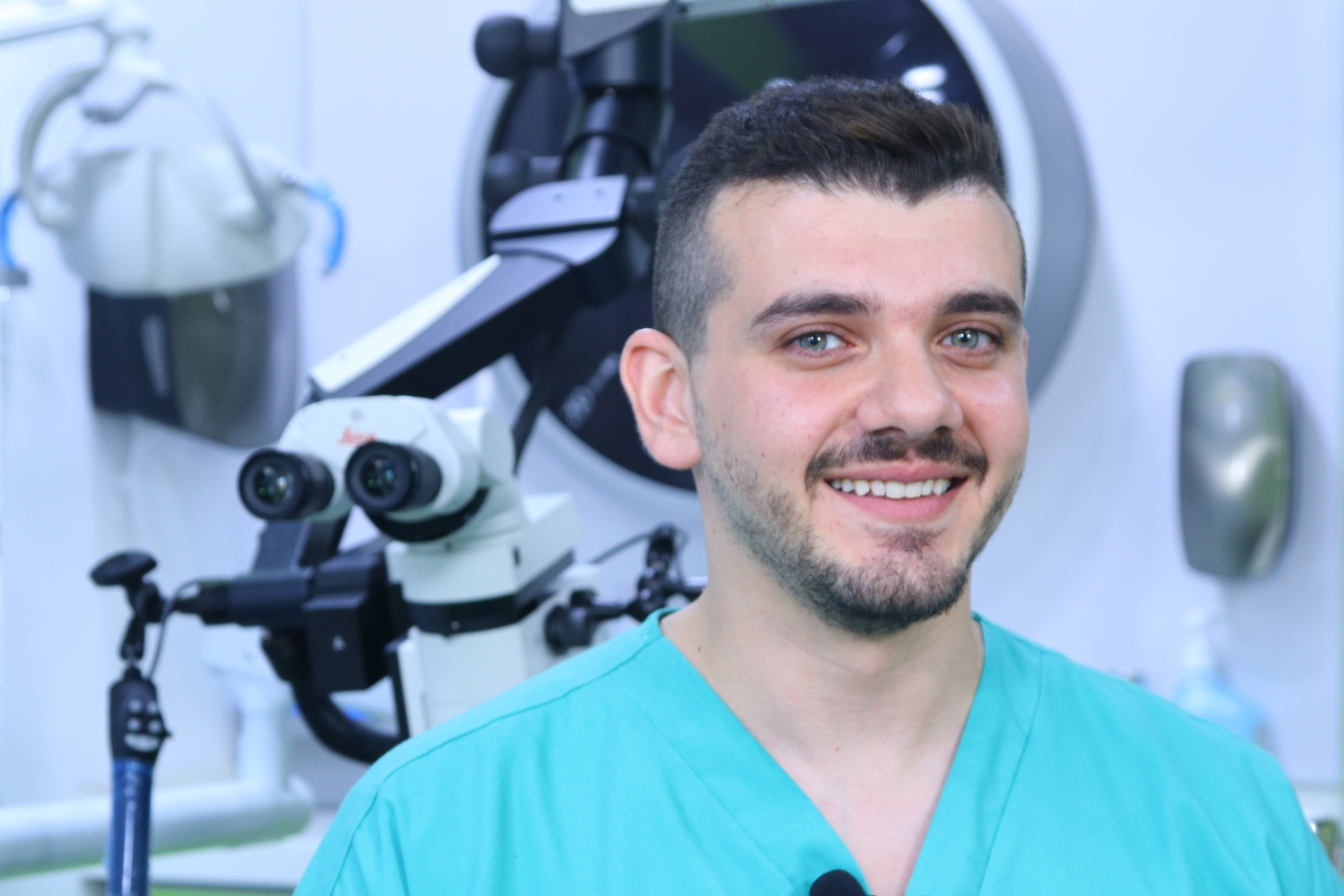
Overcoming Complexities in Microdentistry
Dr. Salam Abu Arqub, from the Smile Engineer Dental Center in Amman, Jordan, has been using Leica dental microscopes for three years for all procedures performed at the clinic. He shared his…
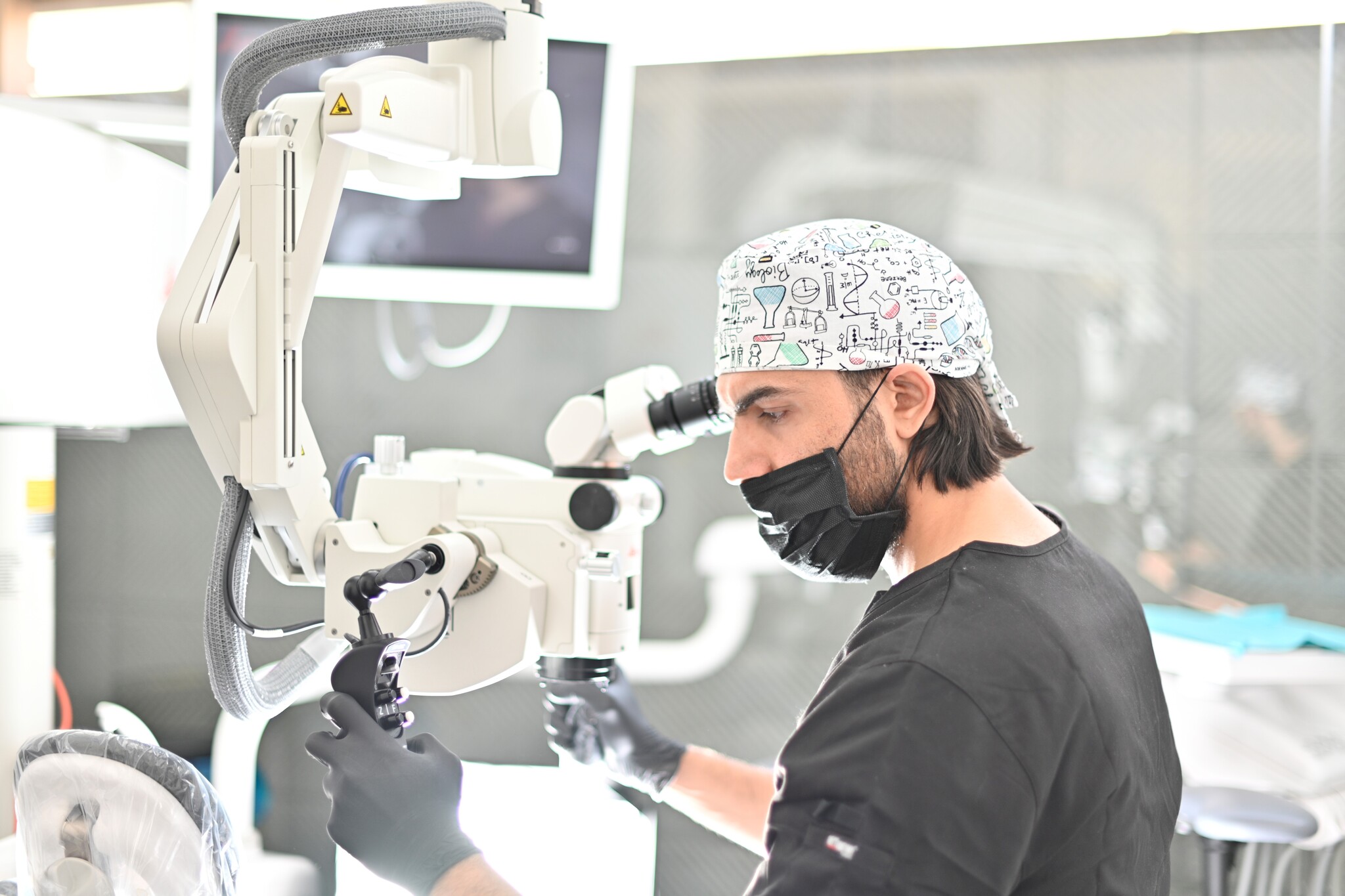
Minimally Invasive Dentistry: Visualization & Posture
Microscopes do not only provide better visualization during dental surgery. They also help ensure correct posture to avoid back pain and neck injuries. Dr. Iyad Ghoneim, from the Safad Dental Center…