メディカル

メディカル

メディカル

脳神経外科、眼科、形成外科、耳鼻咽喉科、歯科の専門医向けに厳選された、最新の科学・臨床リソースを探索しませんか? 貴重な症例や洞察、シンポジウムなど、多彩なコンテンツをご覧いただけます。 手術用顕微鏡の最先端技術に焦点を当て、AR蛍光、3D視覚化、術中OCTイメージングといった革新的技術が、複雑な手術の精度向上や意思決定の最適化にどのように貢献するかをご紹介します。

Fluorescence Recovery after Photobleaching (FRAP) and its Offspring

FRAP (Fluorescence recovery after photobleaching) can be used to study cellular protein dynamics: For visualization the protein of interest is fused to a fluorescent protein or a fluorescent dye. A…

Förster Resonance Energy Transfer (FRET)

The Förster Resonance Energy Transfer (FRET) phenomenon offers techniques that allow studies of interactions in dimensions below the optical resolution limit. FRET describes the transfer of the energy…

An Introduction to CARS Microscopy

CARS overcomes the drawbacks of conventional staining methods by the intrinsic characteristics of the method. CARS does not require labeling because it is highly specific to molecular compounds which…
Scheme of a 2D mosaic scan. Drosophila melanogaster (eye section)

Mosaic Images

Confocal laser scanning microscopes are widely used to create highly resolved 3D images of cells, subcellular structures and even single molecules. Still, an increasing number of scientists are…

Confocal Optical Section Thickness

Confocal microscopes are employed to optically slice comparably thick samples.
Section taste buds rabbit, differential interference contrast microscope

Optical Contrast Methods

Optical contrast methods give the potential to easily examine living and colorless specimens. Different microscopic techniques aim to change phase shifts caused by the interaction of light with the…
Modulation contrast visualizes transparent, low-contrast specimens.

Integrated Modulation Contrast (IMC)

Hoffman modulation contrast has established itself as a standard for the observation of unstained, low-contrast biological specimens. The integration of the modulator in the beam path of themodern…

New Standard in Electrophysiology and Deep Tissue Imaging

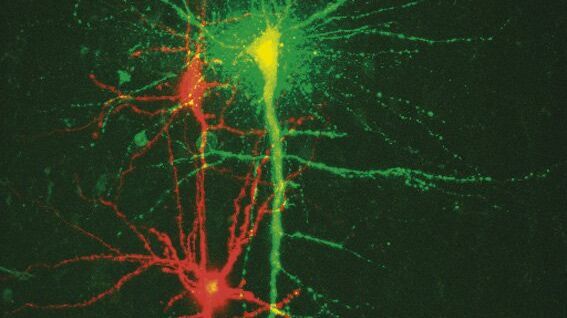
The function of nerve and muscle cells relies on ionic currents flowing through ion channels. These ion channels play a major role in cell physiology. One way to investigate ion channels is to use…
A portion of an early binocular microscope developed by John Leonhard Riddel in the early 1850s.

The History of Stereo Microscopy

This article gives an overview on the history of stereo microscopes. The development and evolution from handcrafted instruments (late 16th to mid-18th century) to mass produced ones the last 150…
Scroll to top