

神経科学研究
神経変性疾患の理解向上に取り組んでいる、もしくは神経系の機能を研究をしていますか? ライカマイクロシステムズのイメージングソリューションによってブレイクスルーを起こす方法をご覧ください。

ゼブラフィッシュを用いた研究
スクリーニング、ソーティング、マニピュレーションおよびイメージングを通じて最良の結果を得るためには、細部や構造を観察して、研究の次の段階に向けて正しい判断を下す必要があります。
優れた光学系と高解像度で定評のあるライカの実体顕微鏡と透過照明スタンドは、世界中の研究者から支持されています。
レーザーマイクロダイセクション
小動物などの解剖を行うとき、顕微鏡の接眼レンズを何時間ものぞくことがあります。 ライカ マイクロシステムズでは、さまざまな顕微鏡と幅広い解剖顕微鏡部品やアクセサリーから選ぶことができるため、ニーズに最適な顕微鏡ソリューションを見つけることができます。

A Guide to Spatial Biology
What is spatial biology, and how can researchers leverage its tools to meet the growing demands of biological questions in the post-omics era? This article provides a brief overview of spatial biology…

神経細胞移動の分子的秘密を解き明かす
発達中の脳における特定部位への神経細胞の移動を調べるには、さまざまなアプローチを用いることができます。このウェビナーでは、オックスフォード大学の専門家が、神経発達過程における大脳皮質の機能層への神経細胞移動の分子メカニズムを解明するために使用している顕微鏡ツールとアッセイについて紹介します。これらのプロセスを理解することは、健全な脳の発達の理解を深め、神経発達障害の治療法を改善する可能性につながり…

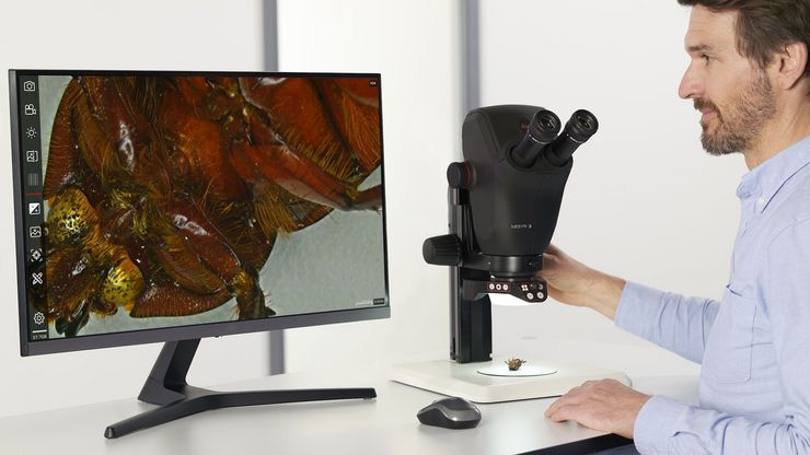
Key Factors to Consider When Selecting a Stereo Microscope
This article explains key factors that help users determine which stereo microscope solution can best meet their needs, depending on the application.
. Image courtesy of Prof. Hui Guo, School of Life Sciences, Central South University, China")
機械受容性経路とシナプス経路の研究に顕微鏡がいかに役立つか
このポッドキャストでは、Tobi Langenhan教授は、顕微鏡を使ってシナプスのタンパク質集合体を調べるなど、接着型GPCRの機械受容特性の研究を通して、タンパク質のダイナミクスとその空間的相互作用に精通されています。 Abdullah…

How to do a Proper Cell Culture Quick Check
In order to successfully work with mammalian cell lines, they must be grown under controlled conditions and require their own specific growth medium. In addition, to guarantee consistency their growth…
 in 14 x 18 tiles. Lifetime gives an additional contrast that allows to differentiate different structures in histological stainings.")
A Guide to Fluorescence Lifetime Imaging Microscopy (FLIM)
The fluorescence lifetime is a measure of how long a fluorophore remains on average in its excited state before returning to the ground state by emitting a fluorescence photon.
