

Accelerating Neuron Image Analysis with Automation
The ability to examine complex neural processes relies on the accurate reconstruction of neuronal networks at scale. Most data extraction methods in neuroscience research are time-consuming and…

Designing your Research Study with Multiplexed IF Imaging
Multiplexed tissue analysis is a powerful technique that allows comparisons of cell-type locations and cell-type interactions within a single fixed tissue sample. It is common for researchers to ask…

Be Confident in your Results with Cell DIVE Validated Antibodies
The Cell DIVE System includes a carefully curated list of hundreds of commercially available antibodies validated to offer optimal specificity and sensitivity in multiplexed imaging. That validation…
 and astrocytes (green) in a cortical spheroid derived from human induced pluripotent stem cells.")
Neuroscience Images
Neuroscience commonly uses microscopy to study the nervous system’s function and understand neurodegenerative diseases.

Understanding Motor Sequence Generation Across Spatiotemporal Scales
We have developed a microscopy-based pipeline to characterize a developmentally critical behavior at the pupal stage of development, called the ecdysis sequence. We study brain-wide neuronal activity…

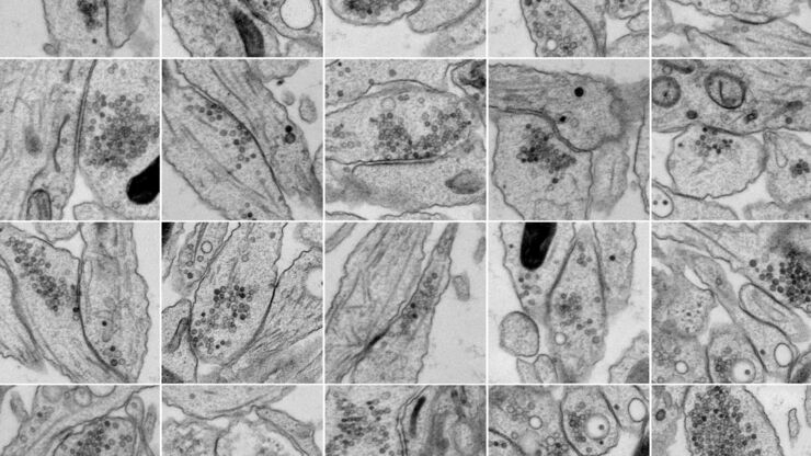
Investigating Synapses in Brain Slices with Enhanced Functional Electron Microscopy
A fundamental question of neuroscience is: what is the relationship between structural and functional properties of synapses? Over the last few decades, electrophysiology has shed light on synaptic…
Bridging Structure and Dynamics at the Nanoscale through Optogenetics and Electrical Stimulation
Nanoscale ultrastructural information is typically obtained by means of static imaging of a fixed and processed specimen. However, this is only a snapshot of one moment within a dynamic system in…

Zebrafish Brain - Whole Organ Imaging at High Resolution
Structural information is key when one seeks to understand complex biological systems, and one of the most complex biological structures is the vertebrate central nervous system. To image a complete…

Ratiometric Imaging
Many fundamental functions of a cell strongly depend on delicate, but nevertheless dynamic balances of ions (e.g. calcium, magnesium), voltage potentials and pH between the cell’s cytosol and the…
