
High contrast images in real-time
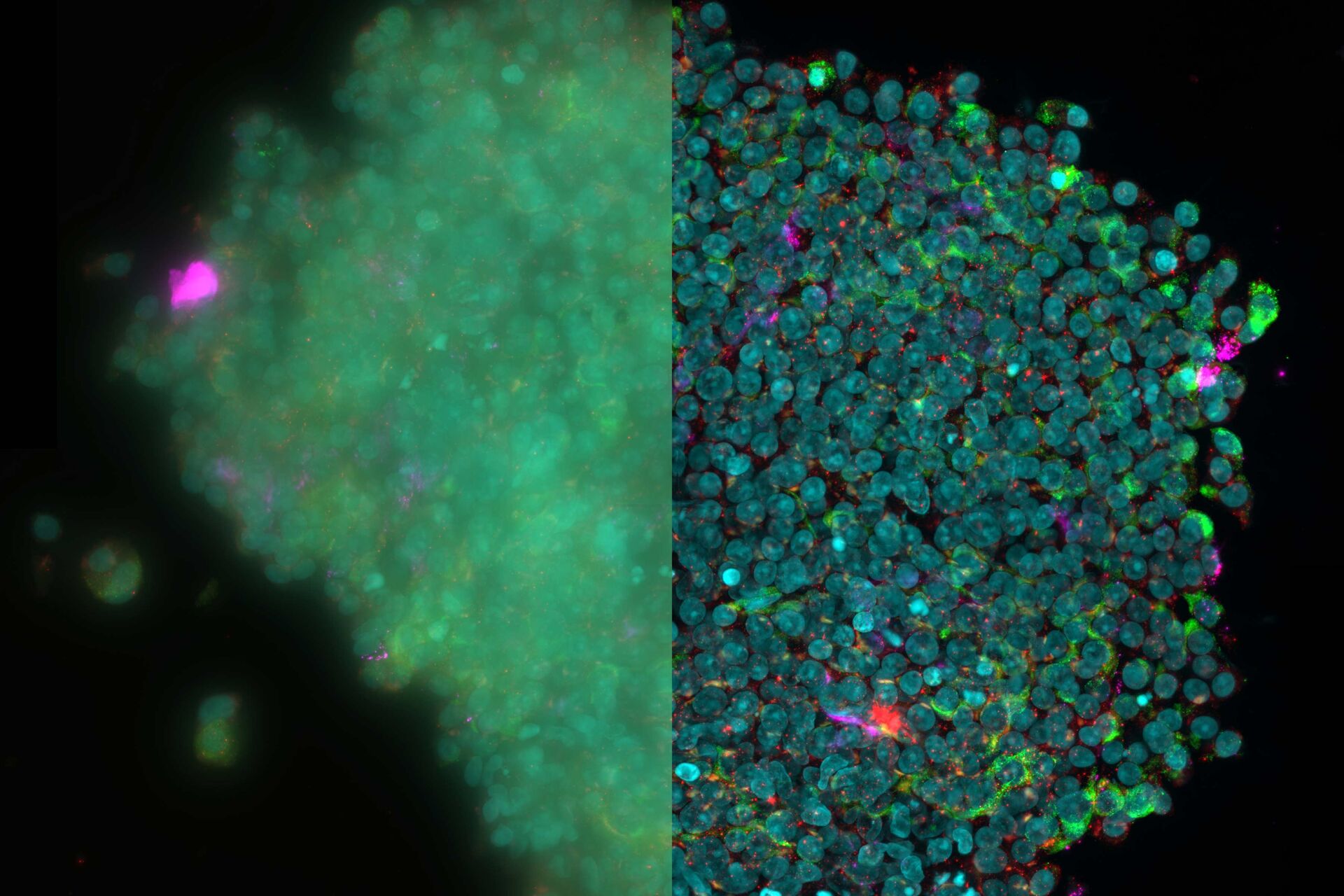
When using conventional methods, imaging with a low light dosage can produce low-quality images with low contrast levels, causing blurred and low-resolution images. This is especially true in thick samples, where out-of-focus blur masks the complexity of the biological structures. THUNDER’s computational clearing approaches are overcoming this challenge by reducing the background on an image in real time, producing crisper images with higher contrast, making even thick samples accessible for high-powered segmentation and analysis.

The real benefit of THUNDER imaging systems is that high image quality does not come at the cost of high light dosage. Gentle widefield imaging avoids damage to samples and ensures their longevity, allowing for multiple scans of large, thick samples over long periods of time. This low-cost high-reward approach maximizes data collected from each experiment, allowing more robust and rapid image analysis of single cells, tissues,
and whole organisms.
Improving image analysis with AI
Aivia is designed to make segmentation and analysis powerful, simple, and reproducible. It uses AI-driven tools like object detection and interpretation to segment and analyze highly complex images. Aivia’s suit of image visualization and analysis packages are designed to automate complex analysis in huge datasets in the range of hundreds of GBs. For example, Aivia can detect and track 2D and 3D objects (like cells, nuclei, organelles, particles, etc.) in time-lapse, in 2-5D reconstructed stacks, and analyze a wide range of morphology, intensity, and motion measurements.
However, accurate segmentation and powerful analysis require high-quality images, which is where combining Aivia and THUNDER comes in. Results are especially powerful when Aivia is combined with computationally cleared THUNDER images, as the high contrast images allow for more rapid and accurate segmentation and analysis. Aivia’s machine learning-based analysis allows lower exposure images to be taken, reducing photo stress on cells.
Overall, combining THUNDER´s high contrast images with Aivia’s AI-driven segmentation and analysis maximizes the physiological relevance of camera-based imaging. The automated, AI-driven package also means that analysis lacks subjective bias, meaning that data can be reproducible across experiments and experimenters.

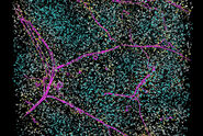
A: THUNDERed and segmented image of the mitochondrial network using Aivia. Different shapes of Mitochondria are visible.
B: Snapshot of a timeseries used to track the mobility, number of objects, size, and the fusion and fission rate of mitochondria over time. The analysis of the THUNDERed dataset shows a larger number of objects, indicating a better accuracy of segmentation. Consequently, the tracking is more precise and any information extracted (displacement, speed, formfactor, size, shape, etc.) is less prone to error.
Conclusion
The use of state-of-the-art AI systems is pushing image analysis into a new generation. Challenges like the conflict between imaging power and sample integrity are being overcome with THUNDER’s computational systems which push image quality and contrast at low light levels. Moreover, AI-based segmentation and analysis with Aivia is enabling researchers to perform ever more powerful and reproducible analysis on high-quality images.
Related Articles
-

A Meta-cancer Analysis of the Tumor Spatial Microenvironment
Learn how clustering analysis of Cell DIVE datasets in Aivia can be used to understand…
Apr 26, 2024Read article -
 tissue.")
Mapping the Landscape of Colorectal Adenocarcinoma with Imaging and AI
Discover deep insights in colon adenocarcinoma and other immuno-oncology realms through the potent…
Apr 26, 2024Read article -

Spatial Architecture of Tumor and Immune Cells in Tumor Tissues
Dig deep into the spatial biology of cancer progression and mouse immune-oncology in this poster,…
Apr 26, 2024Read article
Related Pages
-
Microscope Imaging Software
Microscope imaging software from Leica Microsystems combines microscope, digital camera and…
Visit related page -
THUNDER Imaging Systems
To answer important scientific questions, THUNDER Imaging Systems enable you to obtain a clear view…
Visit related page -
Save Time and Effort with AI-assisted Fluorescence Image Analysis
The powerful synergy of THUNDER and Aivia analyze fluorescence images with greater accuracy, even…
Visit related page