ライフサイエンス

ライフサイエンス

ライフサイエンス

様々な科学分野における顕微鏡の知識、研究技術、そして実用的な応用を深めるための場です。正確な観察、画像解析、そして研究の進歩を実現する方法を学びましょう。高度な顕微鏡技術、イメージング技術、サンプル前処理、画像解析に関する専門的な知見を提供します。最先端のアプリケーションやイノベーションを中心に、細胞生物学、神経科学、がん研究などの分野を幅広くカバーしています。
TIRF Image of Tubulin, YFP, penetration depth: 120 mm

Applications of TIRF Microscopy in Life Science Research

The special feature of TIRF microscopy is the employment of an evanescent field for fluorophore excitation. Unlike standard widefield fluorescence illumination procedures with arc lamps, LEDs or…
TIRF image of brest carcinoma tumor cells expressing GFP tagged cell adhesion Molecule CD44 that is expressed on the cell membrane, imagined in TIRF.

Total Internal Reflection Fluorescence (TIRF) Microscopy

Total internal reflection fluorescence (TIRF) is a special technique in fluorescence microscopy developed by Daniel Axelrod at the University of Michigan, Ann Arbor in the early 1980s. TIRF microscopy…

Super-Resolution GSDIM Microscopy

The nanoscopic technique GSDIM (ground state depletion microscopy followed by individual molecule return) provides a detailed image of the spatial arrangement of proteins and other biomolecules within…
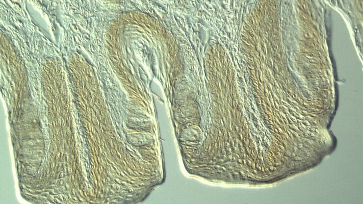
Section taste buds rabbit, differential interference contrast microscope

Optical Contrast Methods

Optical contrast methods give the potential to easily examine living and colorless specimens. Different microscopic techniques aim to change phase shifts caused by the interaction of light with the…
Modulation contrast visualizes transparent, low-contrast specimens.

Integrated Modulation Contrast (IMC)

Hoffman modulation contrast has established itself as a standard for the observation of unstained, low-contrast biological specimens. The integration of the modulator in the beam path of themodern…
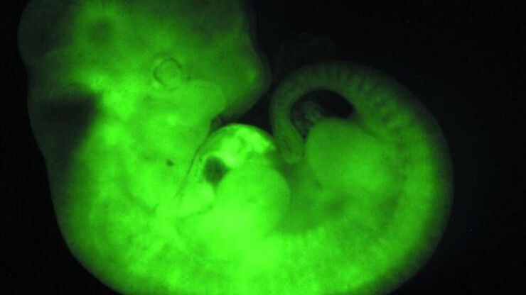
Transgenic Mouse Embryo, GFP

Fluorescence in Microscopy

Fluorescence microscopy is a special form of light microscopy. It uses the ability of fluorochromes to emit light after being excited with light of a certain wavelength. Proteins of interest can be…
Scroll to top