Confocal Microscopes Articles

Super-Resolution Microscopy Image Gallery
Due to the diffraction limit of light, traditional confocal microscopy cannot resolve structures below ~240 nm. Super-resolution microscopy techniques, such as STED, PALM or STORM or some…

Extended Live-cell Imaging at Nanoscale Resolution
Extended live-cell imaging with TauSTED Xtend. Combined spatial and lifetime information allow super-resolution microscopy at extremely low light dose.
 dataset, showing the biochemically distinct structures of a fresh, untreated apple slice.")
How to Prepare Samples for Stimulated Raman Scattering (SRS) imaging
Find here guidelines for how to prepare samples for stimulated Raman scattering (SRS), acquire images, analyze data, and develop suitable workflows. SRS spectroscopic imaging is also known as SRS…

New Imaging Tools for Cryo-Light Microscopy
New cryo-light microscopy techniques like LIGHTNING and TauSense fluorescence lifetime-based tools reveal structures for cryo-electron microscopy.

Find Relevant Specimen Details from Overviews
Switch from searching image by image to seeing the full overview of samples quickly and identifying the important specimen details instantly with confocal microscopy. Use that knowledge to set up…

How to Target Fluorescent Structures in 3D for Cryo-FIB Milling
This article describes the major steps of the cryo-electron tomography workflow including super-resolution cryo-confocal microscopy. We describe how subcellular structures can be precisely located in…
, unsaturated lipids (magenta, 3050 cm-1), collagen (SHG, cyan). Sample courtesy of R. Rudolf, J Klicks, Hochschule Mannheim")
The Potential of Coherent Raman Scattering Microscopy at a Glance
Coherent Raman scattering microscopy (CRS) is a powerful approach for label-free, chemically specific imaging. It is based on the characteristic intrinsic vibrational contrast of molecules in the…

Simplifying Complex Fluorescence Multiwell Plate Assays
Apoptosis, or programmed cell death, occurs during organism embryo development to eliminate unwanted cells and during healing in adults to rid the body of damaged cells and help prevent cancer.…

Efficient Long-term Time-lapse Microscopy
When doing time-lapse microscopy experiments with spheroids, there are certain challenges which can arise. As the experiments can last for several days, prolonged sample survival must be achieved…

Live-Cell Imaging Techniques
The understanding of complex and/or fast cellular dynamics is an important step for exploring biological processes. Therefore, today’s life science research is increasingly focused on dynamic…

Imaging of Anti-Cancer Drug Uptake in Spheroids using DLS
Spheroid 3D cell culture models mimic the physiology and functions of living tissues making them a useful tool to study tumor morphology and screen anti-cancer drugs. The drug AZD2014 is a recognized…
 in 3D")
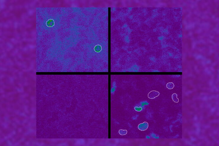
Artificial Intelligence and Confocal Microscopy – What You Need to Know
This list of frequently asked questions provides “hands-on” answers and is a supplement to the introductory article about Dynamic Signal Enhancement powered by Aivia "How Artificial Intelligence…
How Artificial Intelligence Enhances Confocal Imaging
In this article, we show how artificial intelligence (AI) can enhance your imaging experiments. Namely, how Dynamic Signal Enhancement powered by Aivia improves image quality while capturing the…

Benefits of Combining STED and Lifetime
In this interview, Professor Alberto Diaspro talks about the advantages of the White Light Laser and the TauSTED capabilities of STELLARIS 8 STED. He speaks about his experience with the confocal…
Spectroscopic Evaluation of Red Blood Cells
Hemoglobinopathies are a major healthcare problem. This study presents a possible diagnostic tool for thalassemia which is based on confocal spectroscopy. This approach exploits spectral detection and…

The Cryo-CLEM Journey
This article describes the Cryo-CLEM technology and the benefits it can provide for scientists. Additionally, some scientific publications are highlighted.
Recent developments in cryo electron…

Fluorescence Lifetime-based Imaging Gallery
Confocal microscopy relies on the effective excitation of fluorescence probes and the efficient collection of photons emitted from the fluorescence process. One aspect of fluorescence is the emission…

Live Cell Imaging Gallery
Live cell microscopy techniques are fundamental to get a better understanding of cellular and molecular function. Today, widefield microscopy is the most common technique used to visualize cell…
