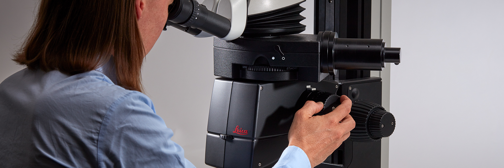
Phase Contrast Light Microscopes
Brightfield microscopy normally only provides a low-contrast image of many transparent biological specimens where few details are distinguished. One way to enhance contrast with brightfield microscopy is to use selective stains, but such stains are often toxic to living cells. A phase contrast light microscope offers a way to view the structures of many types of biological specimens in greater contrast without the need of stains. The contrast method exploits differences in optical density between structures of a specimen that lead to a phase shift of the light that interacts with the specimen and its structures.
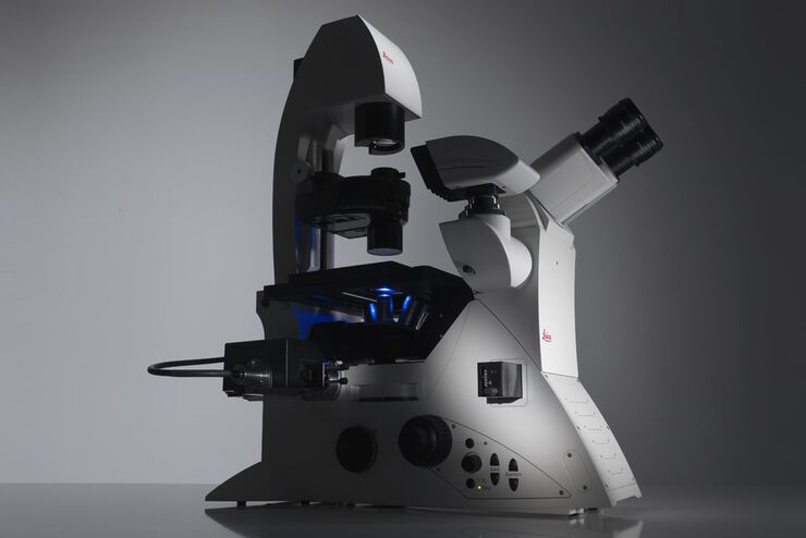
Leica microscopes offer phase contrast for the study of cells or tissues concerning various life-science and forensic applications. Phase contrast can also be useful for certain material and earth-science applications.
Contact us for expert advice on the right phase contrast light microscope for your needs and budget.

What is phase contrast?
Phase contrast is an optical contrast technique for microscopy which makes unstained structures in the cells of biological specimens visible. Cell structures that appear transparent with brightfield illumination can be viewed in high contrast and rich detail using phase contrast. Differences in optical density between structures in the cell can cause light that interacts with them to attain a phase shift. This phenomenon is the basis of phase contrast. As a result, more optically dense structures will look darker than less optically dense ones.

What are some phase contrast microscope uses? Which kind of samples can you visualize?
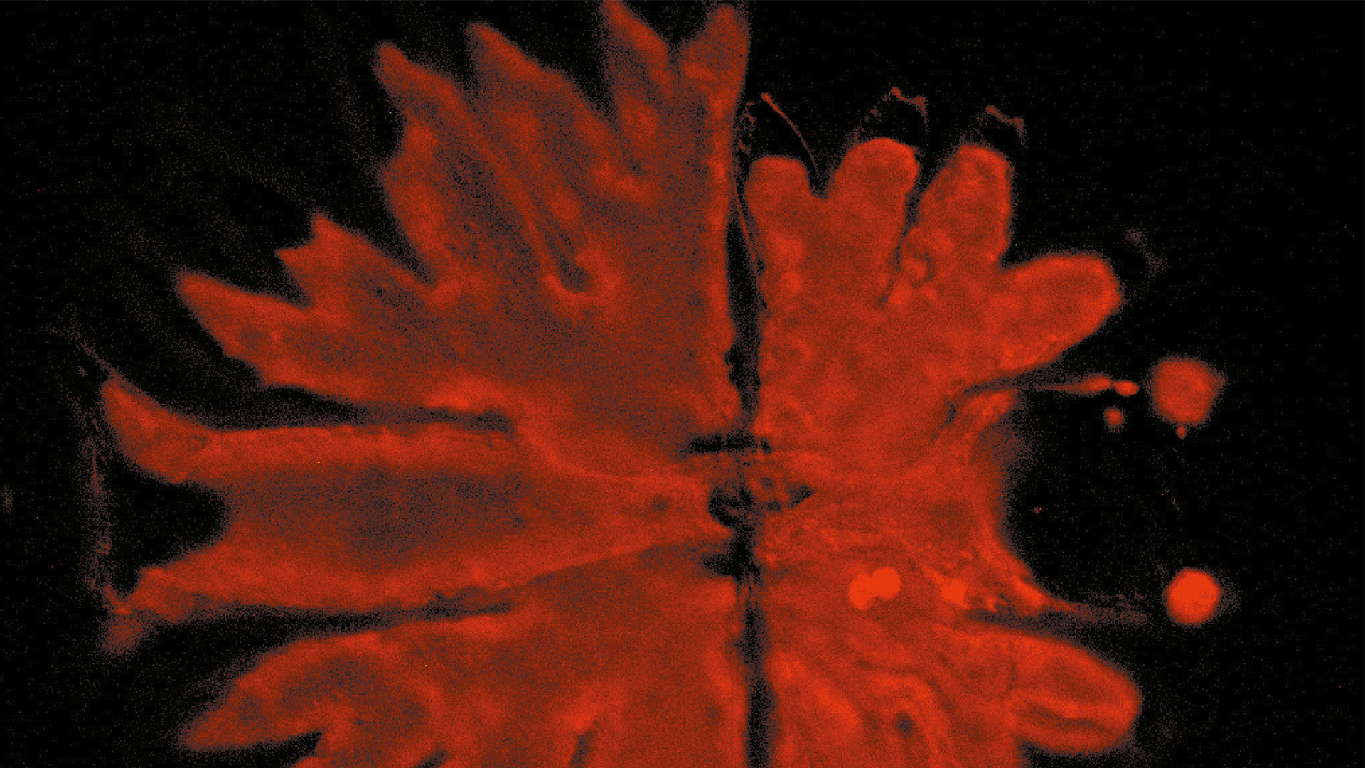
Most often biological specimens and tissues are observed with a phase contrast microscope. A large variety of biological specimens can be observed with phase contrast from fixed specimens to living cells and tissues. For examples, refer to the articles: Phase Contrast & Optical Contrast Methods

How does a phase contrast microscope work?
A phase contrast microscope is similar to a conventional widefield microscope, except it uses an aperture in the shape of an annulus and a quarter-wave (λ/4) phase plate. The annular aperture is placed between the light source and condenser lens and the phase plate after the objective inside the microscope optics. Ring-shaped light that passes through the aperture is focused by the condenser onto the biological specimen to be observed.
Portions of the ring-shaped light are diffracted by optically dense structures of the specimen and experience a negative phase shift of about λ/4. This phase-shifted, diffracted light bypasses the λ/4 plate. In contrast, the portion of the ring-shaped light that passes directly through the specimen non-deviated will hit the phase plate which causes a positive λ/4 phase shift. As the total difference in phase shift between the light diffracted by the specimen’s structures and that which passes through phase plate will be about λ/2, destructive interference will occur. Consequently, more optically dense structures will appear darker than those that are less optically dense.
For more details, refer to the article: Phase Contrast


Using phase shifts for image formation
Frequently Asked Questions Phase Contrast Microscopes
The phase contrast method for microscopy was developed in the 1930s by the Dutch physicist Frits Zernike. After 1942, it became a widely used microscopy technique. In 1953, Zernike was awarded the Nobel Prize for Physics. For more details, refer to the articles: A Brief History of Light Microscopy – From the Medieval Reading Stone to Super-Resolution & Phase Contrast
A phase contrast microscope is similar to a conventional brightfield microscope, except it uses an annular aperture in front of the light source and a quarter-wave phase plate after the objective lens. For more information, refer to the article: Phase Contrast
Yes, a phase contrast microscope can be equipped with a camera for recording images observed with the contrast method.
Related Articles
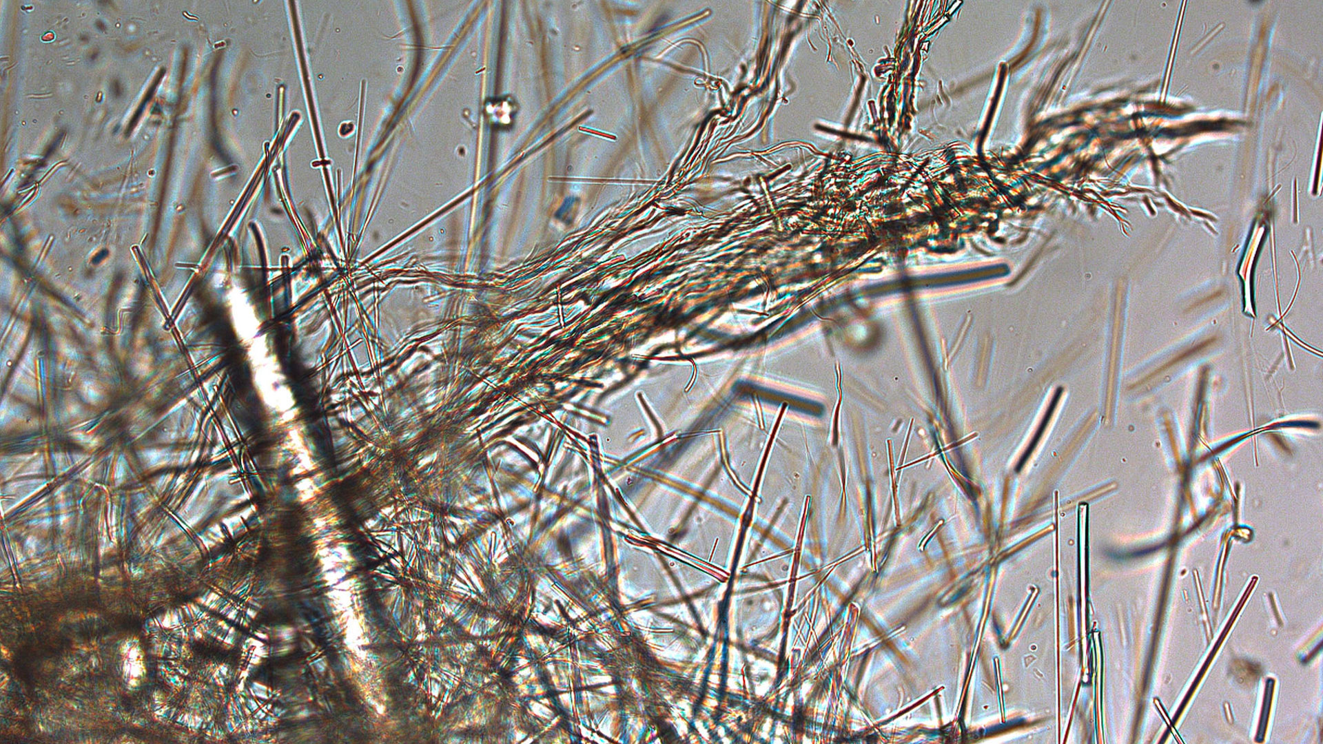
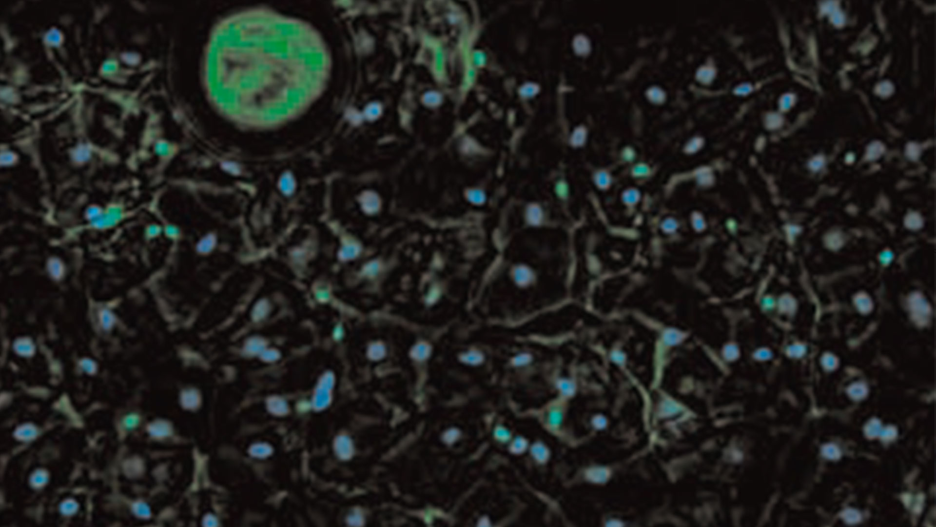
 cells taken with phase contrast.")
Phase Contrast and Microscopy

How to Determine Cell Confluency with a Digital Microscope

How to do a Proper Cell Culture Quick Check
Factors to Consider When Selecting a Research Microscope

Introduction to Mammalian Cell Culture

Introduction to Widefield Microscopy
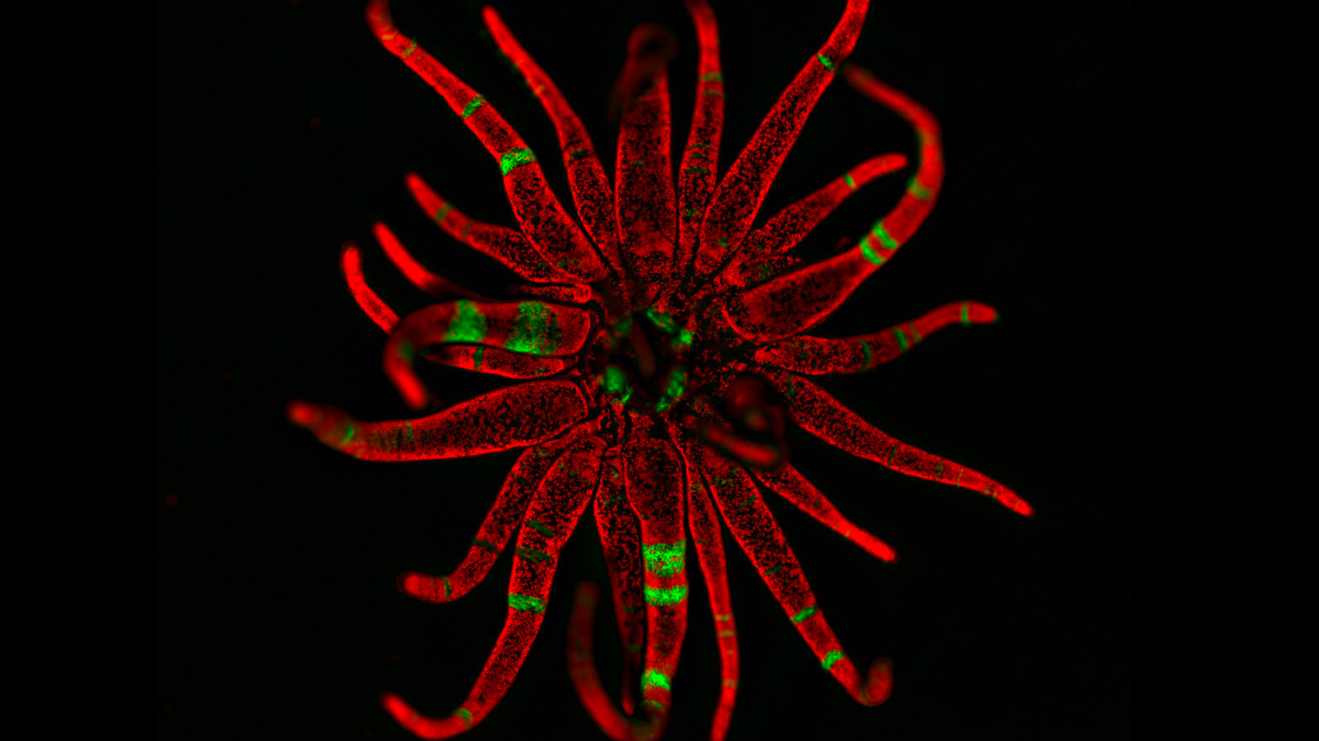
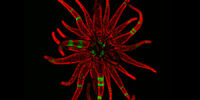
 inoculated with cowpea mosaic virus (CPMV) containing the GFP-gene inserted between the movement protein (MP) and the capsid proteins (CPs) in the viral RNA 2")
Introduction to Live-Cell Imaging

Optical Contrast Methods

Forensic Detection of Sperm from Sexual Assault Evidence
Interested to know more?
Talk to our experts.
Do you prefer personal consulting? Show local contacts