Ion Beam Milling

Ion Beam Milling Systems

When material specimen surfaces are prepared for SEM or incident light microscopy, the specimen usually undergoes multiple processes until the layer or surface to be analyzed is machined with precision. Leica Microsystems’ workflow solutions for solid state technology cover all steps required for demanding high-quality sample preparation.

Need Assistance?

Contact our team of experts to help you simplify your EM sample preparation.

Simplify your EM Sample Preparation with workflow solutions by Leica Microsystems!

While the Leica EM TXP unites all steps of pre-preparation in one instrument, the Leica EM TIC 3X carries out the final, high quality surface finishing for almost any material. Connecting with the Leica EM VCT docking configuration, the sample can then be transferred into the (cryo) SEM under optimal conditions.

Enhanced Resolution with Ion Beam Milling

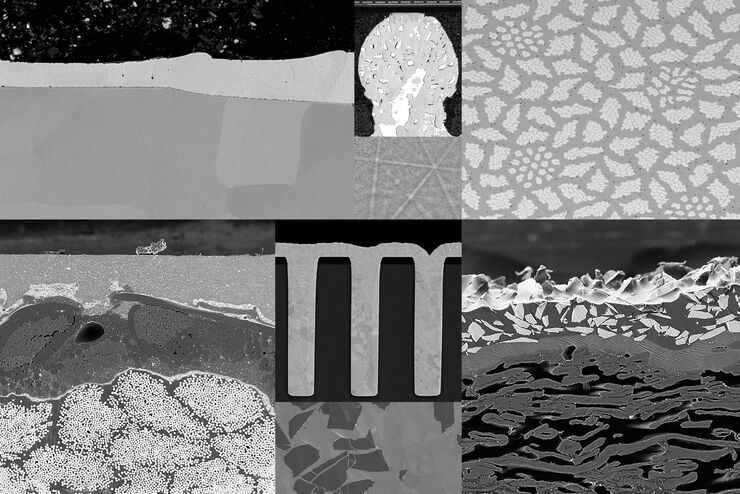
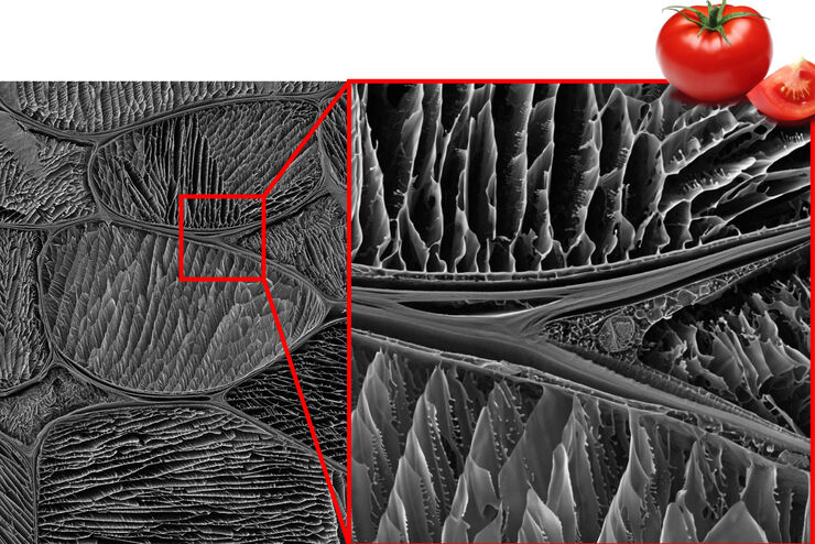
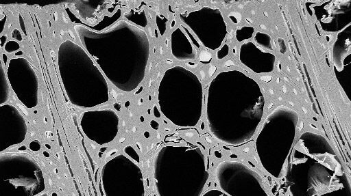
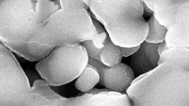
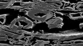
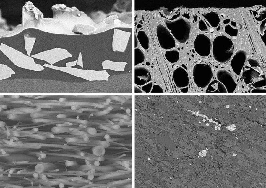
The Ion Beam Milling technique, also known as Ion Beam Etching, is used to achieve a well-prepared sample surface quality for high resolution imaging and analysis. It removes residual artefacts from mechanical cutting and polishing. The ion polished cross-sections and planar samples prepared by Ion Beam Etching can be used for electron microscopy imaging as well as microstructural analysis applications such as EDS, WDS, Auger and EBSD.

The EM TIC 3X Milling Machine offers triple ion beams that speed up the preparation process significantly and achieve to reveal finest details and structures on sample surfaces. Watch the video and see how to cut the preparation time for IC gold wire bonding using the Target Surfacing System EM TXP and the EM TIC 3X Ion Beam Milling System.

ion beam milling related articles

Read our latest articles about Ion Beam Milling

The knowledge portal of Leica Microsystems offers scientific research and teaching material on the subjects of microscopy. The content is designed to support beginners, experienced practitioners and scientists alike in their everyday work and experiments.

More Articles
SEM image of the full Li-NMC electrode sample, showing the two porous layers and the metal film at the center of the structure.

Cross Section Ion Beam Milling of Battery Components

Sample Preparation of Lithium battery systems requires high quality surface preparation to evaluate their internal structure and morphology. Due to the brittle materials involved, preparing pristine…

Introduction to Ion Beam Etching with the EM TIC 3X

In this article you can learn how to optimize the preparation quality of your samples by using the ion beam etching method with the EM TIC 3X ion beam milling machine. A short introduction of the…

Studying the Microstructure of Natural Polymers in Fine Detail

The potential of cryogenic broad ion beam milling used in combination with scanning electron microscopy (cryo-BIB-SEM) for imaging and analyzing the microstructure of cryogenically stabilized soft…

Practical Applications of Broad Ion Beam Milling

Mechanical polishing can be time consuming and frustrating. It can also introduce unwanted artifacts when preparing cross-sectioned samples for electron backscatter diffraction (EBSD) in the scanning…

Porous Ceramics - Sample Preparation for SEM

Application Note for Leica EM RES102 - Ceramic membrane filters with pore sizes down to a few nanometres must be investigated in cross-section with regard to the structure of the pores. The smallest…

Each Atom Counts: Protect Your Samples Prior to FIB Processing

Application Note for Leica EM ACE600 - Focused ion beam (FIB) technology has become an indispensable tool for site-specific TEM sample preparation. It allows to extract electron transparent specimens…

Paper Samples - Sample Preparation for SEM

Application Note for Leica EM RES102 - A coated paper sample has been prepared with ion beam slope cutting in order to test the procedure with regard to its applicability. With the use of ion beam…

Ion Beam Polishing of Sample Surfaces - Sample Preparation for SEM

Application Note for Leica EM RES102 - Ion milling can be used to reduce the roughness of sample surfaces. Small angles less than 6° with respect to the sample surface are necessary. The high voltage…

Triple-beam Ar-Ion-Milling with a Rotary Stage to Decorate Grain Boundaries and Substructures in Rock Salt

Decoration of grain boundaries in polycrystalline rocks has a long tradition in Structural Geology as in a monomineralic rock the recrystallized grain size is a good indicator for the paleostress…

High Quality Sample Preparation for EBSD Analysis by Broad Ion Beam Milling

Electron Backscatter Diffraction technique (EBSD) is known as a "surface" technique because electron diffraction is generated within a few tens of nanometers of the sample surface. Therefore, the…

Cutting and Polishing

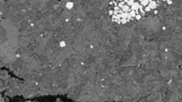
Preparation often begins with a need to accurately cut, grind and polish the surface prior to ion beam milling and metal/carbon coating. With the Leica EM TXP target preparation system all required machining steps can be completed in one instrument – from diamond cutting and milling to polishing.

High quality surfacing

The unique broad ion beam milling system of the Leica EM TIC 3X is the system of choice for EDS, WDS, Auger and EBSD, because ion beam milling is often found to be the only method capable of achieving high quality cross-sections and planed surfaces of almost any material. The process reveals the internal structures of a sample whilst minimizing deformation or damage.

Sputter Coater & Freeze Fracture Solutions

Sputter Coater & Freeze Fracture Solutions

To obtain high-quality images of samples with scanning (SEM) or transmission electron microscopy (TEM), your samples need to be conductive to avoid charging. If a sample does not have a high enough conductivity, then you can quickly cover it with a conductive layer using the method of sputter coating. Also, a carbon or e-beam evaporator coating can be used. Such coatings protect the sample, allow enhancing of the EM image contrast, or can act as a TEM-grid support film for small scale samples.

Read More

 Due to the open chamber design and continuous gas exchange the SampLink chambers keep live cells healthy

Cryo Preparation Systems

High pressure freezing is often the preferred method for preserving aqueous samples in their close-to-native state, as it captures the intricate changes in fine structure or cellular dynamics. Leica Microsystems combines high pressure freezing with light stimulation or electrical stimulation: It enables you to visualize highly dynamic processes or the structural changes of samples at a nanometer resolution and with millisecond precision.

Read More

Overview of Drosophila gastric caeca and a mitochondrion with well-defined cristae and well-preserved surrounding membranes. Micrographs courtesy of Dr. Syed (NIH/NIDCR) and Dr. Bleck (NIH/NHLBI)

Ultramicrotomes & Cryo-Ultramicrotomes

Whether tissue sample, polymer, rubber, metals or nanoparticles, Leica ultramicrotomes provide extremely thin sections and perfect surface quality in a wide range of applications. From materials science to cancer research, our ultramicrotomes are used for many different kinds of research and quality control all over the world. 

Read More

Sputter Coater & Freeze Fracture Solutions
 Due to the open chamber design and continuous gas exchange the SampLink chambers keep live cells healthy
Overview of Drosophila gastric caeca and a mitochondrion with well-defined cristae and well-preserved surrounding membranes. Micrographs courtesy of Dr. Syed (NIH/NIDCR) and Dr. Bleck (NIH/NHLBI)

Interested to know more?

Talk to our experts. We are happy to answer all your questions and concerns.

Contact Us

Do you prefer personal consulting? Show local contacts

Scroll to top