
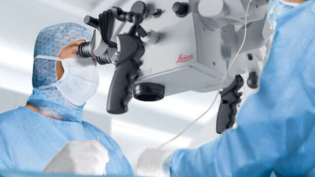

Advanced Visualization in Head and Neck Reconstructive Surgery
PRS surgeons and maxillofacial specialists face unique challenges in reconstructive procedures, particularly when operating in deep, narrow cavities and working with delicate tissues. Achieving…
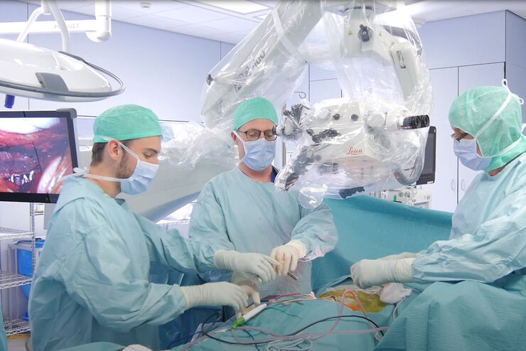
Advanced Visualization: Transforming Minimally Invasive Spine Surgery
Over the past decade, spine surgery has evolved rapidly with the adoption of minimally invasive surgery (MISS) techniques. These approaches reduce surgical trauma, speed up recovery, and help improve…

Advanced Visualization in ENT Surgery: Prof. Darrouzet’s Expert Insights
Otologic surgery demands exceptional precision when working within the confined spaces of the middle and inner ear. Procedures such as tympanoplasty, mastoidectomy, and cochlear implant placement…
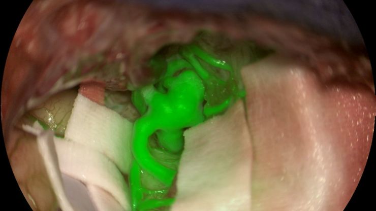
How AR Fluorescence Imaging Supports Neurovascular Surgery
In this article, we explain how fluorescence imaging works in vascular neurosurgery and explain the benefits of the GLOW800 Augmented Reality fluorescence application.
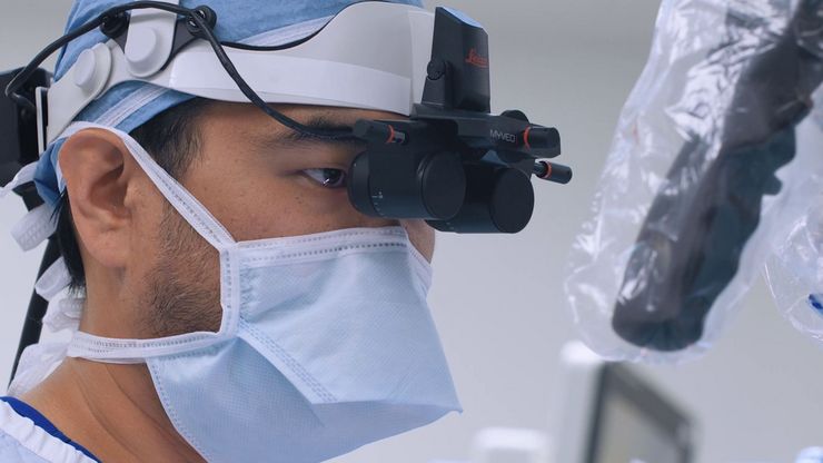
A Microvascular Surgeon’s View: How MyVeo Transforms Visualization
In this article, Dr. Andrew T. Huang, MD, FACS, otolaryngologist and a head and neck reconstructive surgeon, shares how digital 3D surgical visualization with the MyVeo headset from Leica Microsystems…
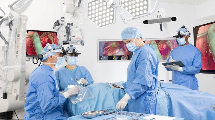
The Guide to Augmented Reality in Microsurgery
In an era of technological advancement, Augmented Reality (AR) is rapidly transforming the medical field. In surgical microscopy, AR can display fluorescence signals as digital overlays in real-time…

Factors to Consider when Selecting Clinical Microscopes
What matters if you would like to purchase a clinical microscope? Learn how to arrive at the best buying decision from our Science Lab Article.
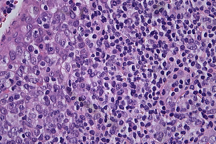
Clinical Microscopy: Considerations on Camera Selection
The need for images in pathology laboratories has significantly increased over the past few years, be it in histopathology, cytology, hematology, clinical microbiology, or other applications. They…
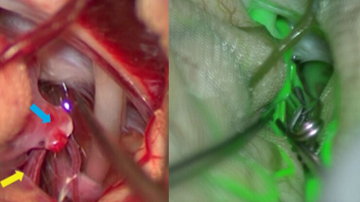
Aneurysm Clipping: Assessing Perforators in Real-Time with AR Fluorescence
This article covers two aneurysm clipping cases highlighting the clinical benefits of GLOW800 Augmented Reality Fluorescence application in neurosurgery, based on insights from Prof. Tohru Mizutani,…


