
Science Lab
Science Lab
Bem-vindo ao portal de conhecimento da Leica Microsystems. Você encontrará pesquisas científicas e material didático sobre o tema microscopia. O portal oferece suporte a iniciantes, profissionais experientes e cientistas em seus trabalhos e experimentos diários. Explore tutoriais interativos e notas de aplicação, descubra os fundamentos da microscopia, bem como as tecnologias de ponta. Faça parte da comunidade do Science Lab e compartilhe sua experiência.
Filter articles
Tags
Story Type
Products
Loading...

Photoactivatable, Photoconvertible, and Photoswitchable Fluorescent Proteins
Fluorescent proteins (FPs) such as GFP, YFP or DsRed are powerful tools to visualize cellular components in living cells. Nevertheless, there are circumstances when classical FPs reach their limits.…
Loading...
 illuminated with wide-band UV excitation. Note the tissue structure with blue autofluorescence. Right: Same tissue and same immunostaining with FITC label illuminated with epi-illumination using narrow")
Milestones in Incident Light Fluorescence Microscopy
Since the middle of the last century, fluorescence microscopy developed into a bio scientific tool with one of the biggest impacts on our understanding of life. Watching cells and proteins with the…
Loading...

Gene Editing with CRISPR/Cas9 - Breakthrough in Genome Engineering
The CRISPR/Cas9 system is one of several different bacterial systems for defense against viral attacks. It consists of two main components. One is a small piece of RNA which binds to the viral target…
Loading...

Studying Caenorhabditis elegans (C. elegans)
Find out how you can image and study C. elegans roundworm model organisms efficiently with a microscope for developmental biology applications from this article.
Loading...

Handbook of Optical Filters for Fluorescence Microscopy
Fluorescence microscopy and other light-based applications require optical filters that have demanding spectral and physical characteristics. Often, these characteristics are application-specific and…
Loading...

Video Tutorial: How to Change the Bulb of a Fluorescence Lamp Housing
When applying fluorescence microscopy in biological applications, a lamp housing with mercury burner is the most common light source. This video tutorial shows how to change the bulb of a traditional…
Loading...

Video Tutorial: How to Align the Bulb of a Fluorescence Lamp Housing
The traditional light source for fluorescence excitation is a fluorescence lamp housing with mercury burner. A prerequisite for achieving bright and homogeneous excitation is the correct centering and…
Loading...
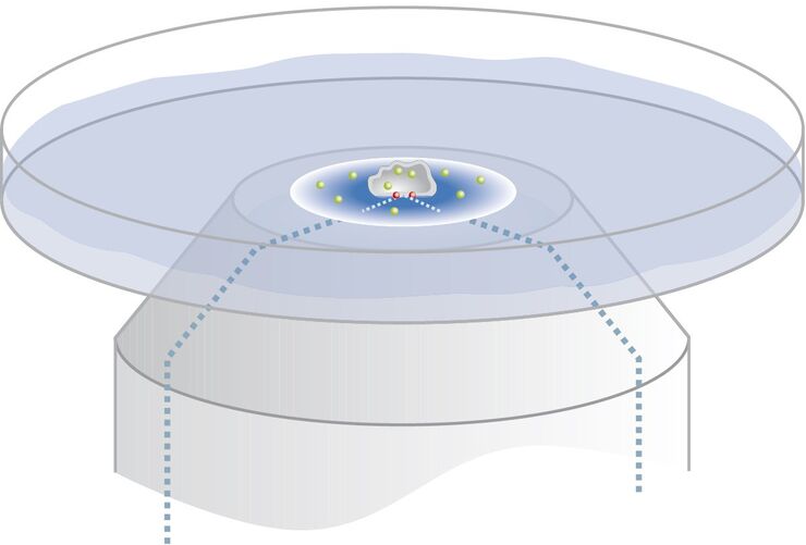
Controlling the TIRF Penetration Depth is Mandatory for Reproducible Results
The main feature of total internal reflection fluorescence (TIRF) microscopy is the employment of an evanescent wave for the excitation of fluorophores instead of using direct light. A property of the…
Loading...

Applications of TIRF Microscopy in Life Science Research
The special feature of TIRF microscopy is the employment of an evanescent field for fluorophore excitation. Unlike standard widefield fluorescence illumination procedures with arc lamps, LEDs or…
