Upright Light Microscopes

Upright microscopes for life science research

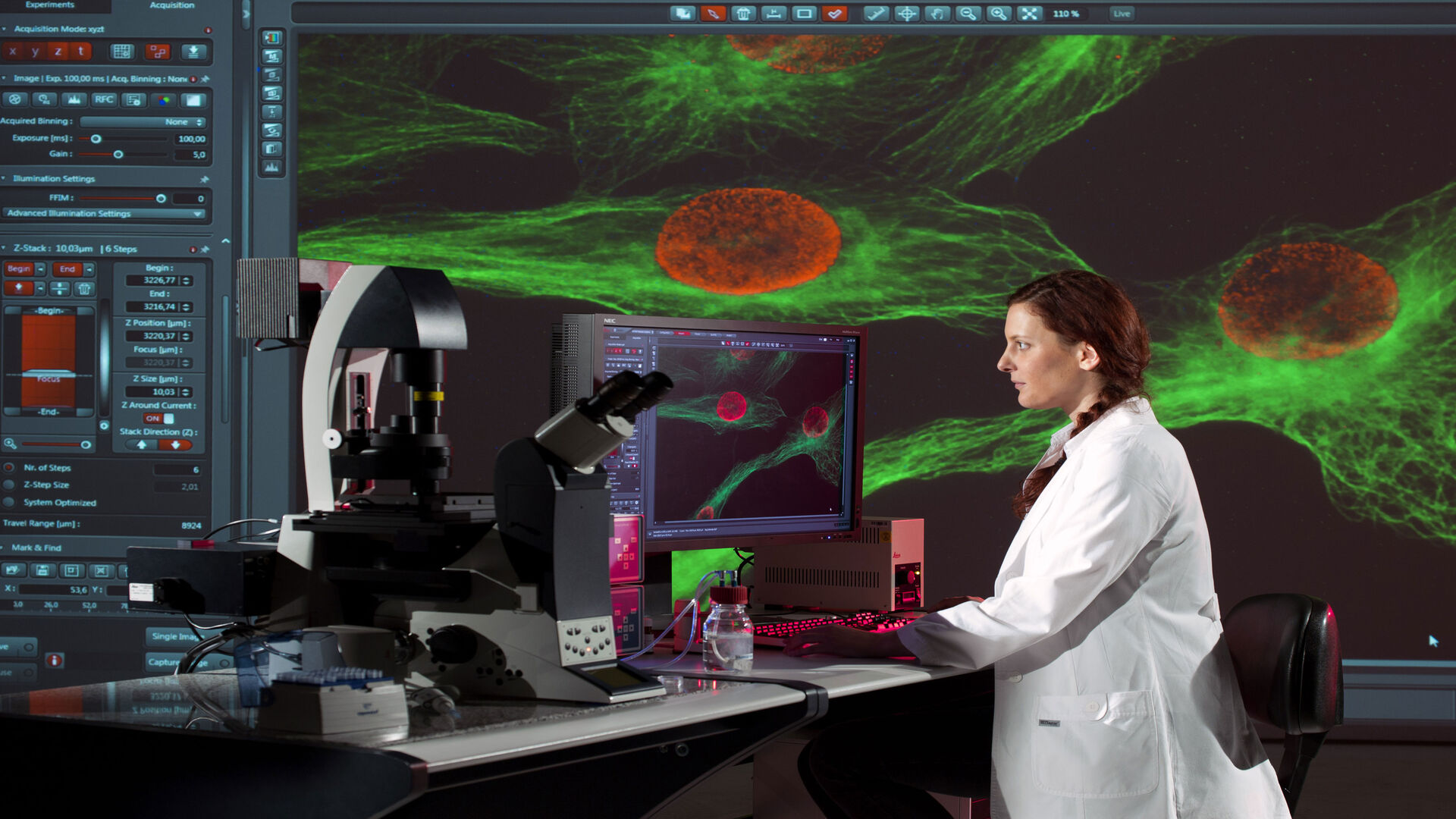
Get the publication-quality imaging and customizable upright microscope solution you need for your Life Science research with Leica Microsystems. These powerful imaging systems feature constant color, natural light illumination, superior optics, and configurable options to provide high contrast, brilliant images for your cutting-edge biological research.

Upright microscopes for industrial and materials

Get insights into the smallest details and inspect and document results efficiently with industrial and materials upright microscopes from Leica Microsystems. Each solution can be customized with brilliant, cool LED illumination, ergonomic accessories, sophisticated digital cameras and intuitive software to meet a broad range of applications.

Need Assistance?

Contact a local imaging specialist for expert advice on the right upright microscope for your needs and budget.

Leica DM4B 6B Upright Microscopes

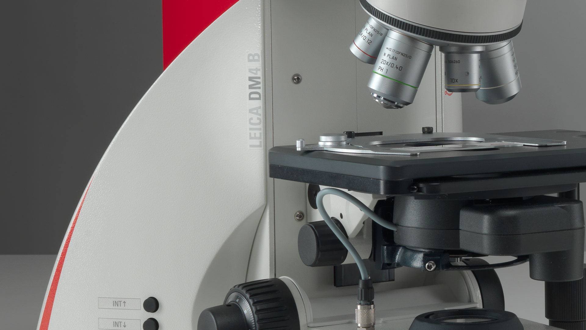
DM4 B & DM6 B Upright Microscopes

Rapid results, customized workflows, and deep insights. Optimized for imaging tissue and plant specimens, as well as pathology, these microscopes are designed to accelerate and tailor your workflows.

DM4 B & DM6 B

Follow us on Instagram

related articles

Read our latest articles about Upright Microscopes

The knowledge portal of Leica Microsystems offers scientific research and teaching material on the subjects of microscopy. The content is designed to support beginners, experienced practitioners and scientists alike in their everyday work and experiments.

More Articles

Some 2D measurements, e.g., lengths and areas, made on a PCB sample with a Leica measurement microscope using the Enersight software.

How to Select the Right Measurement Microscope

With a measurement microscope, users can measure the size and dimensions of sample features in both 2D and 3D, something crucial for inspection, QC, failure analysis, and R&D. However, choosing the…
Example of calibrating a microscope at a higher magnification value using a stage micrometer.

Microscope Calibration for Measurements: Why and How You Should Do It

Microscope calibration ensures accurate and consistent measurements for inspection, quality control (QC), failure analysis, and research and development (R&D). Calibration steps are described in this…
Corneal transplantation. Image courtesy of Mr. David Anderson.

Ophthalmology Case Study: Corneal Transplantation

Learn about the use of intraoperative Optical Coherence Tomography in Corneal Transplantation and how it helps achieve correct positioning of donor tissue.
Optical microscope image, which is a composition of both brightfield and fluorescence illumination, showing organic contamination on a wafer surface. The inset images in the upper left corner show the brightfield image (above) and fluorescence image (below with dark background).

Visualizing Photoresist Residue and Organic Contamination on Wafers

As the scale of integrated circuits (ICs) on semiconductors passes below 10 nm, efficient detection of organic contamination, like photoresist residue, and defects during wafer inspection is becoming…
Image of magnetic steel taken with a 100x objective using Kerr microscopy. The magnetic domains in the grains appear in the image with lighter and darker patterns. A few domains are marked with red arrows. Courtesy of Florian Lang-Melzian, Robert Bosch GmbH, Germany.

Rapidly Visualizing Magnetic Domains in Steel with Kerr Microscopy

The rotation of polarized light after interaction with magnetic domains in a material, known as the Kerr effect, enables the investigation of magnetized samples with Kerr microscopy. It allows rapid…
Region of a patterned wafer inspected using optical microscopy and automated and reproducible DIC (differential interference contrast). With DIC users are able to visualize small height differences on the wafer surface more easily.

6-Inch Wafer Inspection Microscope for Reliably Observing Small Height Differences

A 6-inch wafer inspection microscope with automated and reproducible DIC (differential interference contrast) imaging, no matter the skill level of users, is described in this article. Manufacturing…
Dr. Sheybani places the new subconjunctival stent in the anterior chamber. Images courtesy of Arsham Sheybani, MD.

Glaucoma Stent Revision Surgery Guided by Intraoperative OCT

Learn about a glaucoma subconjunctival stent revision guided by intraoperative OCT and the important role it plays to ensure the best outcome.
Automated wafer loader using carbon fiber end-effectors for safer handling.

Safe Wafer Loading for Microscope Inspection without Hand Contact

How automated silicon wafer loading for microscope inspection helps improve microelectronics process control and production efficiency is explained in this article. Manual handling of wafers has a…
Image of burrs (red arrows) at the edge of a battery electrode acquired with a DVM6 digital microscope.

Burr Detection During Battery Manufacturing

See how optical microscopy can be used for burr detection on battery electrodes and determination of damage potential to achieve rapid and reliable quality control during battery manufacturing.
Plastic reconstructive surgery with M530 OHX

Advances in Oncological Reconstructive Surgery

Decision making and patient care in oncological reconstructive surgery have considerably evolved in recent years. New surgical assistance technologies are helping surgeons push the boundaries of what…

Materials Science and Inspection

Excellent sample preparation and imaging methods are key for inspecting materials and visualizing the fine details with reliability and accuracy. Inverted microscopes enable you to achieve this goal in a more efficient way compared to upright microscopes. They offer you high-quality optics and intelligent automation for optimal workflows. 

Read More

Life Science Research

Inverted microscopes are often used in life science research for the visualization, measurement, analysis, and documentation of live cell and tissue cultures. Continuous innovative technical advances for inverted microscopes support the ever-changing imaging needs of life science researchers. 

Read More

Scroll to top