Alzheimer protein analysis workflow with automatic plaque identification and excision
Leica's Laser Microdissection systems will improve your workflow by automatically identifying and dissecting your ROIs.
This delivers sufficient and pure starting material for proteomics easily. Everything under visual control and without contamination by surrounding tissue.
1. Sample preparation
Typically, special membrane-based slides are used for laser microdissection. Sample preparation for LMD is straightforward, and can be derived from classical preparations for histology.
For downstream proteomics we recommend PET membrane slides (which are almost free of softeners) or DIRECTOR™ slides which allow completely membrane-free ablation.
Paraffin embedding brings the tissue sample into a constitution which can be cut into thin slices. Alternatively you can utilize cryo-sectioning to prepare your slides.
We will deliver the suitable LMD slides for your application.

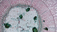
2. Fixation and staining
Amyloid plaques can be visualized by DAB staining based on a β-amyloid antibody. This results in a brown staining of amyloid plaques in brightfield microscopy (right). DAB can be also combined with cresyl violet staining.
An alternative is Congo red which stains amyloid on formalin-fixed, paraffin-embedded (FFPE) as well as frozen tissue sections.
β-amyloid antibodies can also be used for immunofluorescence microscopy which is also applicable with Leica LMD systems.

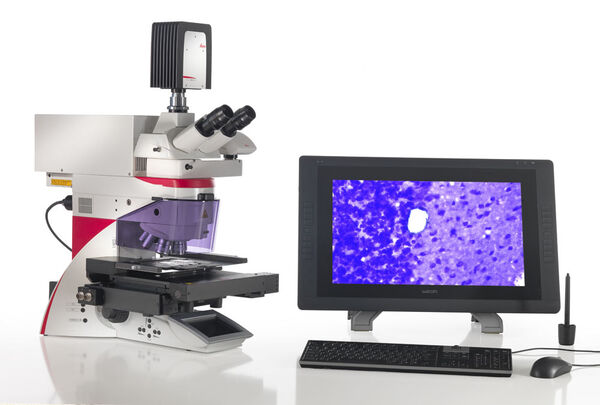
3. Visualization and ROI

Leica LMD systems can generate an automatic sample overview that can be used to easily navigate to your regions of interest (ROI).
Your ROIs can be identified and marked automatically by the ADM software module, or you can apply shapes manually.
- More information on the Leica LMD systems
- Laser microdissection articles & tutorials

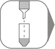
4. Laser microdissection

Next, the defined ROIs are precisely dissected by the laser under visual control and directly collected via gravity.
Isolate disease affected material or any other ROI with a few mouse clicks and pool as much as you want in one or different reaction vessels if desired.
Also, you can cut your samples directly on the fly with the "Move and Cut" tool, without any predefined shapes.
The collection via gravity allows the use of standard, cost-effective consumables such as PCR tubes or 8-strip tubes.

5. Extraction of Proteins
Proteins can be extracted and prepared for downstream analysis from the dissectates with established protocols. Those might differ depending on the upstream sample embedding method, as well as on the aimed protein analysis technique.
Please find some examples here:

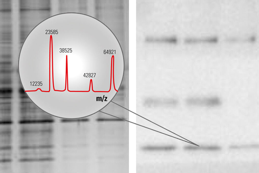
6. Protein expression analysis
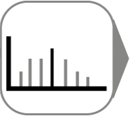
The following techniques for protein analysis have been successfully tested with proteins gained from LMD:
- LC-MS/MS
- ESI-MS/MS
- SDS-PAGE
- Western Blotting
- 2D-DIGE
- MALDI-TOF
- SELDI-TOF
- NMR

Typical fields of research
- Amyloidosis
- Cancer
- Other diseases
- Drug discovery
- Plant research
Register now to download protocol for 'LMD of Brain Tissue for Proteomics'
References
- Drummond et al. Laser Capture Microdissection (2018) pp 319-334
- Hondius et al. Laser Capture Microdissection (2018) pp 371-383
- Cahill et al. RapComMS (2018)
- Drummond et al. ActaNeuropathol 2017
Related Articles
-
.")
Neuron Isolation in Spatial Context with Laser Microdissection (LMD)
After Alzheimer’s disease, Parkinson’s is the second most common progressive neurodegenerative…
Jun 24, 2024Read article -

How did Laser Microdissection enable Pioneering Neuroscience Research?
Dr. Marta Paterlini, a Senior Scientist at the Karolinska Institute, shares her experience of using…
Jun 24, 2024Read article -

Laser Microdissection Protocols for Tissue and Cell Isolation - Download free eBook
In this Bio-protocol Selections, we present a collection of open-access, detailed methods papers…
May 22, 2024Read article
Related Pages
-
Laser Capture Microdissection
Laser Microdissection, also known as LMD or LCM (Laser Capture Microdissection), is a contact- and…
Visit related page