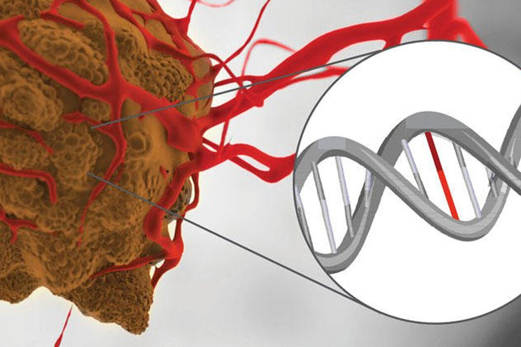
DNA mutation analysis workflow with single cell precision
Leica's Laser Microdissection systems will improve your workflow by precisely cutting only the cells you are interested in. Every step is under visual control and proceeds without contamination with surrounding tissue.
DNA Analysis Workflow - Steps
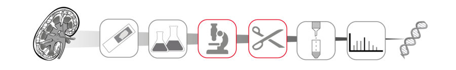
1. Sample preparation
Typically, special membrane-based slides are used for laser microdissection. Sample preparation for LMD is straightforward, and can be derived from classical preparations for histology.
Paraffin embedding brings the tissue sample into a constitution which can be cut into thin slices. Alternatively you can utilize cryo-sectioning to prepare your slides. If fresh cancerous tissue is available, you should prefer cryo-sectioning to preserve nucleic acids from degradation.
We will deliver the suitable LMD slides for your application.
2. Fixation and staining
In general paraffin sections or frozen sections can be used. Here we will outline the use of frozen sections.
Sample slides should be fixed and stained right after cryo-sectioning with acetone, ethanol or a mixture of ethanol and acetic acid.
H&E is a common histology stain, where Hematoxylin stains the nuclei, and eosin stains the cytoplasm.
H&E staining is suitable for both paraffin and frozen sections.

3. Visualization and ROI definition

Leica LMD systems can generate an automatic sample overview that can be used to easily navigate to your regions of interest (ROI).
Your ROIs can be identified and marked automatically by the AVC software module, or you can apply shapes manually.
- More information on the Leica LMD systems
- More information on the Leica LMD application software
- Laser microdissection articles & tutorials

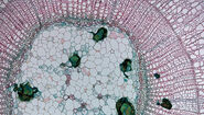
4. Laser microdissection

Next, the defined ROIs are precisely dissected by the laser under visual control and directly collected via gravity. Visually controlled excision prevents mixing tumor material with healthy cells which distort the result. The collection via gravity allows for the use of standard, cost-effective consumables such as PCR tubes or 8-strip tubes.
Also, you can cut your samples directly on the fly with the "Move and Cut" tool, without any predefined shapes.
We provide the appropriate LMD system according to your needs.

5. Extraction of DNA
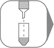
Leica Microsystems recommends the high-quality QIAGEN kits (QIAamp® DNA Micro Kit) for preparation of nucleic acids.
They can be immediately used in downstream applications such as PCR, sequencing, quantitative, real-time PCR, or can be stored at –20°C until needed.
Please refer to www.qiagen.com for details.
6. DNA mutation analysis
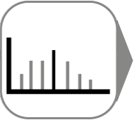
DNA mutation analysis is done via next generation sequencing (NGS).
Other technologies like Pyrosequencing or classical DNA Sanger sequencing are applicable as well.
Typical fields of research
- Pathology research
- Neuroscience
- Pharmaceutical research
- Plant research
- Epigenetics research
Further information
References
- Shen et al., Nature, 2018, doi: 10.1038/s41586-018-0703-0
- Makohon-Moore et al., Nature, 2018, doi: 10.1038/s41586-018-0481-8
- Untch et al., CancerRes, 2018, doi: 10.1158/0008-5472.CAN-17-1925
- Huang et al. Nanotheranostics (2018)
Related Articles
-
.")
Neuron Isolation in Spatial Context with Laser Microdissection (LMD)
After Alzheimer’s disease, Parkinson’s is the second most common progressive neurodegenerative…
Jun 24, 2024Read article -

How did Laser Microdissection enable Pioneering Neuroscience Research?
Dr. Marta Paterlini, a Senior Scientist at the Karolinska Institute, shares her experience of using…
Jun 24, 2024Read article -

Laser Microdissection Protocols for Tissue and Cell Isolation - Download free eBook
In this Bio-protocol Selections, we present a collection of open-access, detailed methods papers…
May 22, 2024Read article
Related Pages
-
Laser Capture Microdissection
Laser Microdissection, also known as LMD or LCM (Laser Capture Microdissection), is a contact- and…
Visit related page