
Science Lab
Science Lab
Bienvenido al portal de conocimiento de Leica Microsystems. Aquí encontrará investigación científica y material didáctico sobre el tema de la microscopía. El portal ayuda a principiantes, profesionales experimentados y científicos por igual en su trabajo diario y en sus experimentos. Explore tutoriales interactivos y notas de aplicación, descubra los fundamentos de la microscopía, así como las tecnologías de gama alta. Forme parte de la comunidad Science Lab y comparta sus conocimientos.
Filter articles
Tags
Story Type
Products
Loading...

Coherent Raman Scattering Microscopy Publication List
CRS (Coherent Raman Scattering) microscopy is an umbrella term for label-free methods that image biological structures by exploiting the characteristic, intrinsic vibrational contrast of their…
Loading...
 dataset, showing the biochemically distinct structures of a fresh, untreated apple slice.")
How to Prepare Samples for Stimulated Raman Scattering (SRS) imaging
Find here guidelines for how to prepare samples for stimulated Raman scattering (SRS), acquire images, analyze data, and develop suitable workflows. SRS spectroscopic imaging is also known as SRS…
Loading...
, unsaturated lipids (magenta, 3050 cm-1), collagen (SHG, cyan). Sample courtesy of R. Rudolf, J Klicks, Hochschule Mannheim")
The Potential of Coherent Raman Scattering Microscopy at a Glance
Coherent Raman scattering microscopy (CRS) is a powerful approach for label-free, chemically specific imaging. It is based on the characteristic intrinsic vibrational contrast of molecules in the…
Loading...

Formulated Product Characterization with SRS Microscopy
Creams, pastes, gels, emulsions, and tablets are ubiquitous across a wide range of manufacturing sectors from pharmaceuticals and consumer health products to agrochemicals and paint. To improve…
Loading...
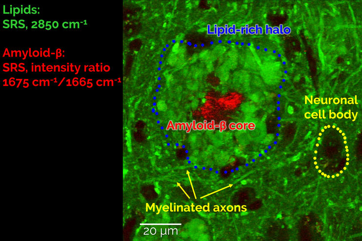
Stimulated Raman Scattering Microscopy Probes Neurodegenerative Disease
Despite decades of research, the molecular mechanisms underlying some of the most severe neurodegenerative diseases, such as Alzheimer’s or Parkinson’s, remain poorly understood. The progression of…
Loading...

CARS Microscopy: Imaging Characteristic Vibrational Contrast of Molecules
Coherent anti-Stokes Raman scattering (CARS) microscopy is a technique that generates images based on the vibrational signatures of molecules. This imaging methods does not require labeling, yet…
Loading...
An Introduction to CARS Microscopy
CARS overcomes the drawbacks of conventional staining methods by the intrinsic characteristics of the method. CARS does not require labeling because it is highly specific to molecular compounds which…
