
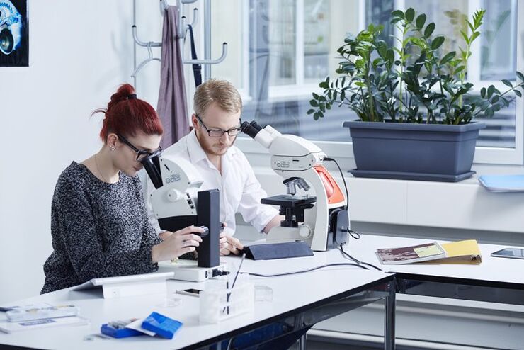
Science Lab
Science Lab
Benvenuti nel portale delle conoscenze di Leica Microsystems. Troverete materiale didattico e di ricerca scientifica sul tema della microscopia. Il portale supporta i principianti, i professionisti esperti e gli scienziati nel loro lavoro quotidiano e negli esperimenti. Esplorate i tutorial interattivi e le note applicative, scoprite le basi della microscopia e le tecnologie di punta. Entrate a far parte della comunità di Science Lab e condividete la vostra esperienza.
Filter articles
Tag
Tipo di storia
Prodotti
Loading...

Organismi Modello nella Ricerca
Un organismo modello è una specie utilizzata dai ricercatori per studiare specifici processi biologici. Hanno caratteristiche genetiche simili a quelle umane e vengono normalmente impiegate in aree di…
Loading...

Microscopy in Virology
The coronavirus SARS-CoV-2, causing the Covid-19 disease effects our world in all aspects. Research to find immunization and treatment methods, in other words to fight this virus, gained highest…
Loading...

Ricerca sul cancro
Il cancro è una malattia complessa ed eterogenea causata da cellule che hanno dei difetti nella regolazione della crescita. I cambiamenti genetici ed epigenetici in una cellula o in un gruppo di…
Loading...
Digital Classroom Options
As teachers, you know your big challenge is to catch and keep the students’ attention and the best chance for this is by making the environment interactive. In the case of the Microscopy Classroom, we…
Loading...
 images")
How To Create EDOF (Extended Depth of Focus) Images
Watch this video to see how you can rapidly record sharp optical microscope images of samples with a large height variation. This is done with the optional Extended Depth of Focus (EDOF) function of…
Loading...
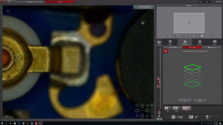
How to Make a Fast Z-stack
Save time for your 2D and 3D analysis. Watch this video to learn about the new user interface, LAS X.next, for the DVM6 digital microscope. The video demonstrates how to make a fast Z-Stack with a few…
Loading...

Digital Microscopy in Earth Science
Classical polarized light (compound) microscopes can only be used for prepared samples, because the working distance they offer is insufficient for whole samples. This means that thicker and bigger…
Loading...

Introduction to Mammalian Cell Culture
Mammalian cell culture is one of the basic pillars of life sciences. Without the ability to grow cells in the lab, the fast progress in disciplines like cell biology, immunology, or cancer research…
Loading...

Introduction to Widefield Microscopy
This article gives an introduction to widefield microscopy, one of the most basic and commonly used microscopy techniques. It also shows the basic differences between widefield and confocal…
