Specimen preparation for LMD using a cryotome
Specimen preparation for laser microdissection is a bit more tricky than for usual microscopy: The content of the tissue needs to be protected from degradation and alteration. In addition, the used fixatives and stains should not influence or effect the downstream analysis. Therefore fresh frozen tissue is the first choice especially for RNA downstream analysis: The fresh material provides highest starting quality and special protocols for fixation. Stain procedures will keep this high RNA quality during the whole upstream process and the UV laser microdissection process has no impact on the RNA quality as well.

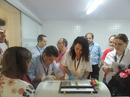
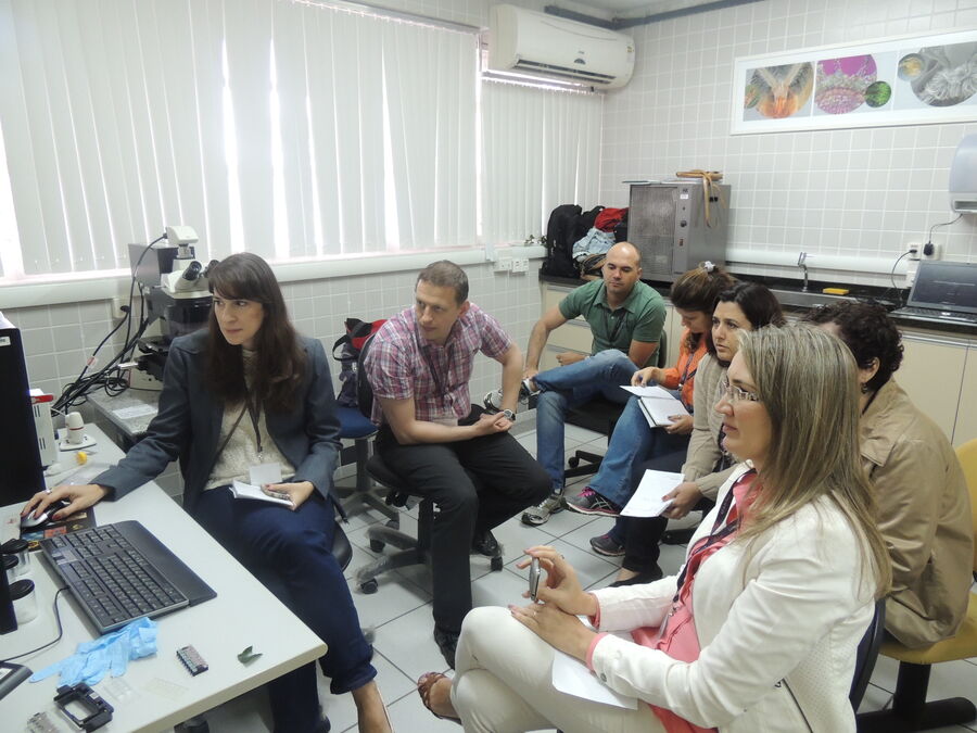
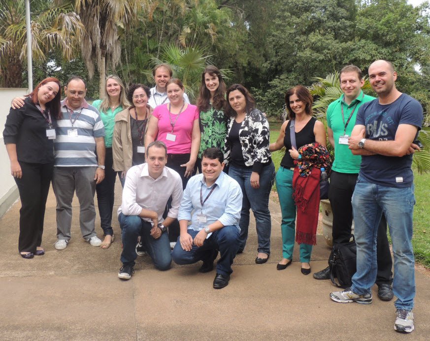
Fig. 1, 2: Dr. Falk Schlaudraff, Product Manager at Leica Microsystems (center, left), explained how to use a Leica cryotome for Laser microdissection sample preparation of freshly frozen tissue.
Chromosome Spread preparation for LMD
Beside tissue preparation and dissection for RNA downstream analysis, DNA analysis (e.g. for mutation detection) is desired. As a single cell commonly contains two pairs of chromosomes, the isolation of single chromosomes is particularly important for chromosome-specific diseases as well as for the understanding of gene structures and contribution of genes in different species.
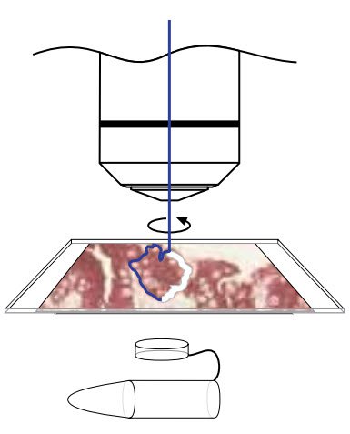
With the Leica LMD system and the 150x dry objective in combination with POL frame slides, individual chromosome isolation is very easy. Step 1: chromosome spread on the POL frame slide needs to be prepared. The Leica LMD protocol guide provides a protocol for such a sample preparation of white blood cells from human blood (kindly provided by Dr. Henry Dijkman, Department of Pathology, Radboud University Nijmegen Medical Center, The Netherlands).
Protocol Steps for a chromosome spread preparation for laser microdissection:
- Cells were cultured for 72 to 96 hours.
- Culture was treated with colcemid for 1 hour.
- Culture was treated with a hypotonic solution for 10-12 minutes.
- Washing step and cell fixation in methanol/acetic acid (3 + 1, v/v).
- Fixed cells were dropped on the POL FrameSlide and air dried overnight.
- Giemsa staining solution was applied for 3 minutes.
- After another washing step in water, slides were air-dried.
- Slides were mounted to the Leica LMD system and a specimen overview in low magnification was created for easier identification of the spreads.
- 150x objective used for a detailed visualization of a spread.
- Clean the surrounding area of the chromosome of interest (Move+Cut or Draw+Scan mode).
- Select a collection device for the chromosome of interest and dissect with suitable laser settings.
This short video shows a new LMD user who practices how to dissect chromosomes according the guidance of the Leica specialists:
The tricky work begins after the collection of the single chromosome, as analysis and amplification of such little material is not easy. However, commercial kits like the Qiagen Repli-G kit can help to manage this challenge. "As proven during this event, the collection of the chromosome itself is feasible and easy to learn," Falk says. "In addition working with the Leica LMD system is fun as it provides much more than looking at slides."
The Leica LMD system is the connecting link between microscopy and molecular biology. "Once you found the correct settings for your system you are ready to go," Falk explains. "And it is worth investing the time to find the best settings for your sample as you can easily store and recover those settings making your life much easier and save valuable time for more success!"
Related Articles
-
.")
Neuron Isolation in Spatial Context with Laser Microdissection (LMD)
After Alzheimer’s disease, Parkinson’s is the second most common progressive neurodegenerative…
Jun 24, 2024Read article -

How did Laser Microdissection enable Pioneering Neuroscience Research?
Dr. Marta Paterlini, a Senior Scientist at the Karolinska Institute, shares her experience of using…
Jun 24, 2024Read article -

Laser Microdissection Protocols for Tissue and Cell Isolation - Download free eBook
In this Bio-protocol Selections, we present a collection of open-access, detailed methods papers…
May 22, 2024Read article