Industrie
Industrie
Tauchen Sie ein in detaillierte Artikel und Webinare, die sich mit effizienter Inspektion, optimierten Arbeitsabläufen und ergonomischem Komfort in industriellen und pathologischen Umgebungen befassen. Zu den behandelten Themen gehören Qualitätskontrolle, Materialanalyse, Mikroskopie in der Pathologie und vieles mehr. Sie erhalten wertvolle Einblicke in den Einsatz von Spitzentechnologien zur Verbesserung der Präzision und Effizienz von Fertigungsprozessen sowie zur präzisen pathologischen Diagnose und Forschung.
Unlocking the Secrets of Organoid Models in Biomedical Research
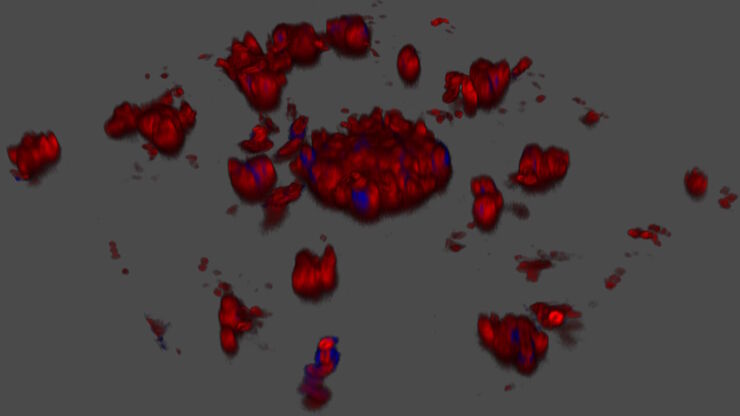
Get ready to delve deeper into the world of organoids and 3D models, which are essential tools for advancing our understanding of human health. Navigating these complex structures and obtaining clear…
Mica: A Game-changer for Collaborative Research at Imperial College London
This interview highlights the transformative impact of Mica at Imperial College London. Scientists explain how Mica has been a game-changer, expanding research possibilities and facilitating…
From Bench to Beam: A Complete Correlative Cryo Light Microscopy Workflow
In the webinar entitled "A Multimodal Vitreous Crusade, a Cryo Correlative Workflow from Bench to Beam" a team of experts discusses the exciting world of correlative workflows for structural biology…
Overcoming Challenges with Microscopy when Imaging Moving Zebrafish Larvae
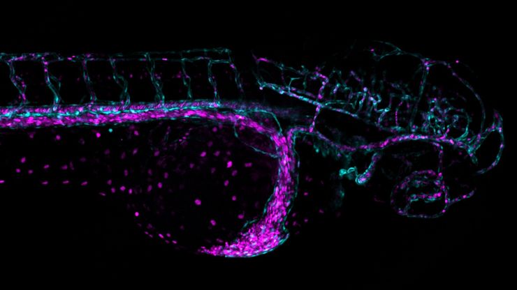
Zebrafish is a valuable model organism with many beneficial traits. However, imaging a full organism poses challenges as it is not stationary. Here, this case study shows how zebrafish larvae can be…
Leitfaden zur Räumlichen Biologie
What is spatial biology, and how can researchers leverage its tools to meet the growing demands of biological questions in the post-omics era? This article provides a brief overview of spatial biology…
Cutting-Edge Imaging Techniques for GPCR Signaling
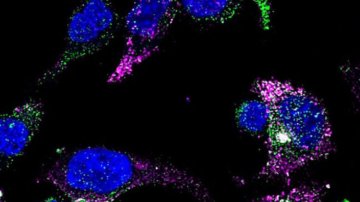
With this webinar on-demand enhance your pharmacological research with our webinar on GPCR signaling and explore cutting-edge imaging techniques that aim to understand how GPCR signaling translates…
Mikrobielle Welten erforschen: Räumliche Interaktionen in 3D Lebensmittelmatrizen
Das Micalis Institute ist eine gemeinsame Forschungseinheit in Zusammenarbeit mit INRAE, AgroParisTech und der Université Paris-Saclay. Seine Mission ist es, innovative Forschung im Bereich der…
How do Cells Talk to Each Other During Neurodevelopment?
Professor Silvia Capello presents her group’s research on cellular crosstalk in neurodevelopmental disorders, using models such as cerebral organoids and assembloids.
Super-Resolution Microscopy Image Gallery
Due to the diffraction limit of light, traditional confocal microscopy cannot resolve structures below ~240 nm. Super-resolution microscopy techniques, such as STED, PALM or STORM or some…


