Science Lab

Science Lab

Science Lab

Benvenuti nel portale delle conoscenze di Leica Microsystems. Troverete materiale didattico e di ricerca scientifica sul tema della microscopia. Il portale supporta i principianti, i professionisti esperti e gli scienziati nel loro lavoro quotidiano e negli esperimenti. Esplorate i tutorial interattivi e le note applicative, scoprite le basi della microscopia e le tecnologie di punta. Entrate a far parte della comunità di Science Lab e condividete la vostra esperienza.

Factors to Consider When Selecting a Research Microscope

An optical microscope is often one of the central devices in a life-science research lab. It can be used for various applications which shed light on many scientific questions. Thereby the…
Influenca in lung epithelial cells (porcine) - THUNDER Imager 3D Cell Culture Influenca virus – red, cilia – green, Nuclei – blue.

How Can Immunofluorescence Aid Virology Research?

Modern virology research has become as crucial now as ever before due to the global COVID-19 pandemic. There are many powerful technologies and assays that virologists can apply to their research into…

Virologia

L’oggetto della tua ricerca si concentra su infezioni e malattie virali? Scopri in che modo puoi approfondire l’aspetto virologico utilizzando le soluzioni di imaging e la preparazione del campione di…

Computational Clearing - Enhance 3D Specimen Imaging

This webinar is designed to clarify crucial specifications that contribute to THUNDER Imagers' transformative visualization of 3D samples and improvements within a researcher's imaging-related…

THUNDER Imagers: High Performance, Versatility and Ease-of-Use for your Everyday Imaging Workflows

This webinar will showcase the versatility and performance of THUNDER Imagers in many different life science applications: from counting nuclei in retina sections and RNA molecules in cancer tissue…

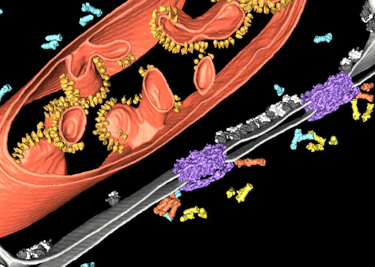
Improve Cryo Electron Tomography Workflow

Leica Microsystems and Thermo Fisher Scientific have collaborated to create a fully integrated cryo-tomography workflow that responds to these research needs: Reveal cellular mechanisms at…

Alzheimer Plaques: fast Visualization in Thick Sections

More than 60% of all diagnosed cases of dementia are attributed to Alzheimer’s disease. Typical of this disease are histological alterations in the brain tissue. So far, there is no cure for this…
HeLa cell spheroid stained with Alexa Fluor 568 Phalloidin (Actin) and YOYO 1 iodide (Nucleus).

Real Time Images of 3D Specimens with Sharp Contrast Free of Haze

THUNDER Imagers deliver in real time images of 3D specimens with sharp contrast, free of the haze or out-of-focus blur typical of widefield systems. They can even image clearly places deep inside a…
Scroll to top