THUNDER Imaging Systems

THUNDER Imaging Systems

THUNDER Imaging Systems

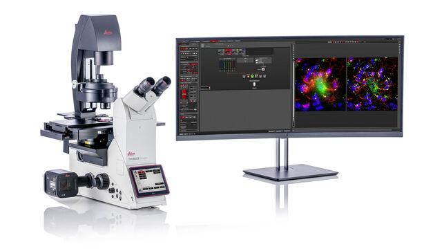
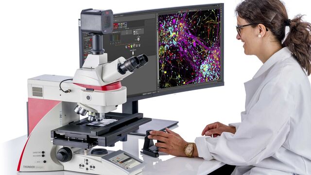
To answer important scientific questions, THUNDER Imaging Systems enable you to obtain a clear view of details, even deep within an intact sample, in real time without out-of-focus blur. THUNDER brings you high-speed, multicolor imaging of thin and thick samples with increased temporal resolution in the first attempt itself. Sharp imaging of 2D and 3D specimens is as easy as working with your favorite camera-based fluorescence microscope.

THUNDER Imaging Systems

THUNDER offers speed and sensitivity. See fine details clearly throughout single cells, tissues, whole organisms, or tumor spheroids.

THUNDER Imager Cell

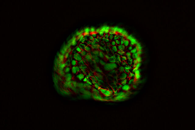
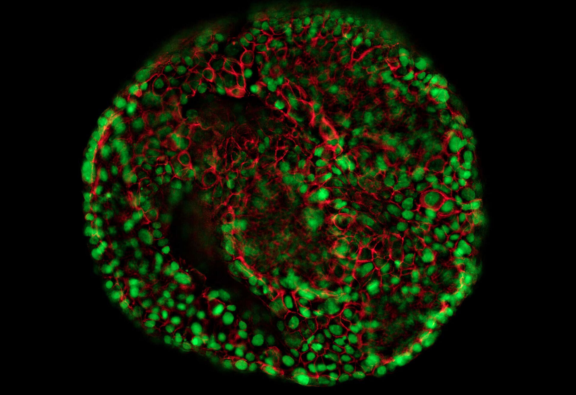
THUNDER Imagers provide you with a solution for advanced 3D cell culture assays, whether you want to study stem cells, spheroids, or organoids.
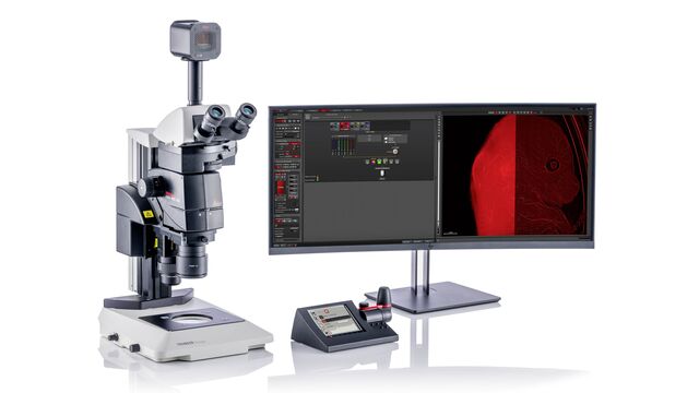
THUNDER Imager Model Organism

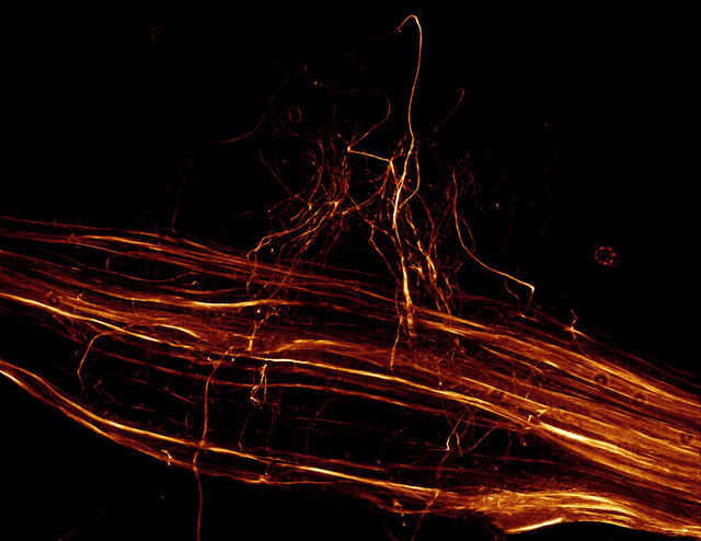
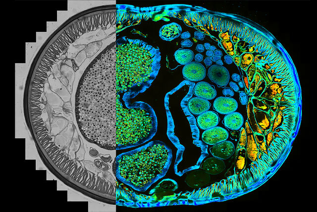
The THUNDER Imager Model Organism allows fast and easy 3D exploration of whole organisms for developmental or molecular biology research.
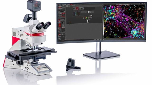
THUNDER Imager Tissue

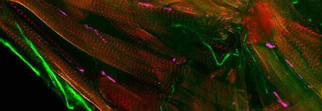
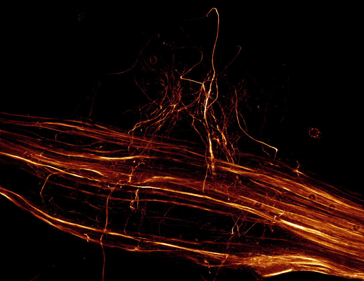
The THUNDER Imager Tissue allows real-time fluorescence imaging of 3D tissue sections typically used in neuroscience and histology research.

Read our latest articles about THUNDER Imaging Systems

The knowledge portal of Leica Microsystems offers scientific research and teaching material on the subjects of microscopy. The content is designed to support beginners, experienced practitioners and scientists alike in their everyday work and experiments.
More Articles

What’s the Best Organoid Imaging Approach for Early Drug Discovery?
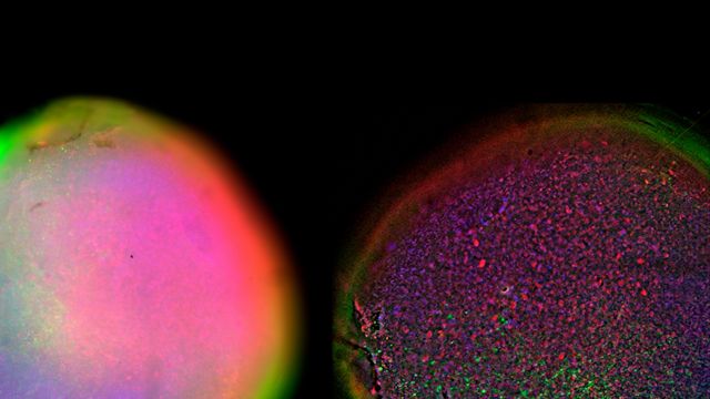
Organoids and other complex in vitro models (CIVMs) are becoming increasingly important in early drug discovery and translational research, driven by the need for more predictive, human-relevant data in line with evolving regulatory expectations. However, scientists face many challenges when imaging and analyzing these 3D systems. This eBook explores how complementary imaging and analysis workflows can help you extract deeper insights from complex 3D biological models.

Fast, High-Contrast Widefield Imaging of Optically Challenging Samples
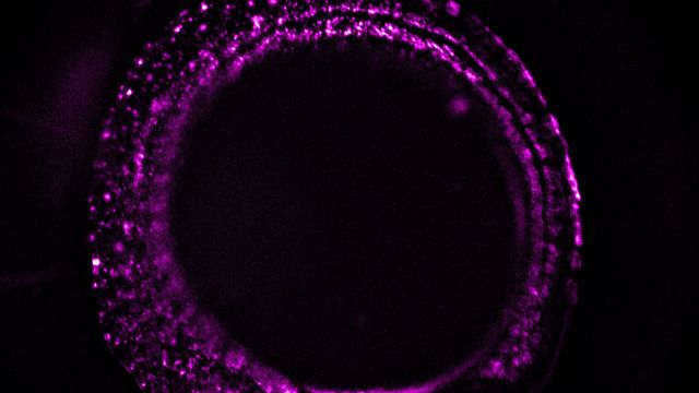
Live‑cell imaging of large, complex biological samples often requires large fields of view, sub-cellular resolution, high-sensitivity, and fast acquisition – all while maintaining low illumination doses for gentle long-term observation. Widefield fluorescence microscopy can meet these demands, but signals can be buried in its inherent out of focus blur. Optical sectioning microscopy techniques can mitigate this, but can introduce trade-offs in speed, phototoxicity, or experimental complexity.

Factors to Consider When Selecting a Research Microscope
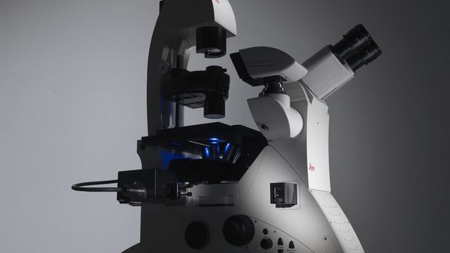
An optical microscope is often one of the central devices in a life-science research lab. It can be used for various applications which shed light on many scientific questions. Thereby the configuration and features of the microscope are crucial for its application coverage, ranging from brightfield through fluorescence microscopy to live-cell imaging. This article provides a brief overview of the relevant microscope features and wraps up the key questions one should consider when selecting a research microscope.

A Guide to C. elegans Research – Working with Nematodes
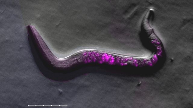
Efficient microscopy techniques for C. elegans research are outlined in this guide. As a widely used model organism with about 70% gene homology to humans, the nematode Caenorhabditis elegans (also called a roundworm), is ideal for studying development, neuroscience, genetics, and aging. Its transparency and ease of cultivation make it an outstanding model system for genetics. It can be imaged at high resolution. Key lab methods include worm picking, transgenesis, fluorescence screening, imaging, and documentation.

Improving Zebrafish-Embryo Screening with Fast, High-Contrast Imaging

Discover from this article how screening of transgenic zebrafish embryos is boosted with high-speed, high-contrast imaging using the DM6 B microscope, ensuring accurate targeting for developmental biology research.

Unlocking the Secrets of Organoid Models in Biomedical Research
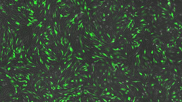
Get ready to delve deeper into the world of organoids and 3D models, which are essential tools for advancing our understanding of human health. Navigating these complex structures and obtaining clear images for analysis can be challenging. In this event, researchers from The University of Oxford and University College London will join us to show how the THUNDER Imager Cell Spinning Disk can provide more convincing, high-quality data for deeper insights into a variety of models.

Designing the Future with Novel and Scalable Stem Cell Culture
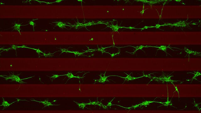
Visionary biotech start-up Uncommon Bio is tackling one of the world’s biggest health challenges: food sustainability. In this webinar, Stem Cell Scientist Samuel East shows how they make stem cell culture media for cellular agriculture safe and economically viable. See how they achieved a 1000x reduction in media costs and developed animal free, food safe iPSC culture media.

Revealing Neuronal Migration’s Molecular Secrets
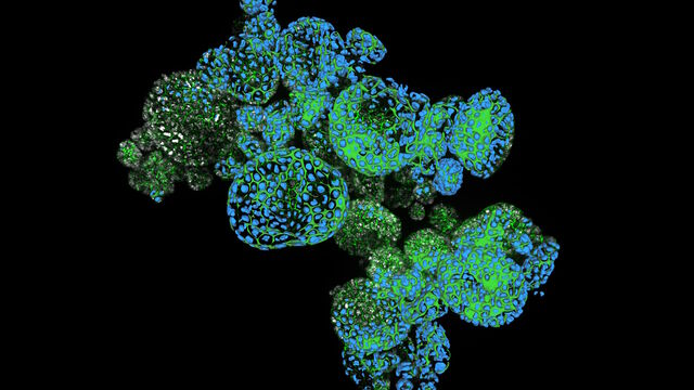
Different approaches can be used to investigate neuronal migration to their niche in the developing brain. In this webinar, experts from The University of Oxford present the microscopy tools and assays they use to elucidate the molecular mechanisms of neuronal migration to functional layers of the cortex during neurodevelopment. Understanding these processes will lead to a better understanding of healthy brain development and potentially better treatments for neurodevelopmental disorders.

How Efficient is your 3D Organoid Imaging and Analysis Workflow?
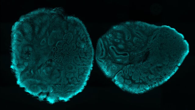
Organoid models have transformed life science research but optimizing image analysis protocols remains a key challenge. This webinar explores a streamlined workflow for organoid research, starting with real-time 3D cell culture checks. This is followed by high-speed, high-resolution 3D imaging to generate crisp images and cleaner data for accurate AI-based segmentation and quantification of parameters such as growth rate, cell migration and 3D cellular interactions – enabling deeper insights.

Imaging Organoid Models to Investigate Brain Health
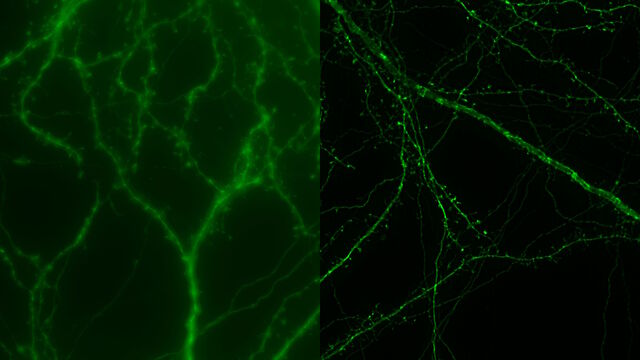
Imaging human brain organoid models to study the phenotypes of specialized brain cells called microglia, and the potential applications of these organoid models in health and disease.

How Microscopy Helps the Study of Mechanoceptive and Synaptic Pathways
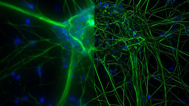
In this podcast, Dr Langenhan explains how microscopy helps his team to study mechanoceptive and synaptic pathways, their challenges, and how they overcome them.

What are the Challenges in Neuroscience Microscopy?
eBook outlining the visualization of the nervous system using different types of microscopy techniques and methods to address questions in neuroscience.

The Role of Iron Metabolism in Cancer Progression
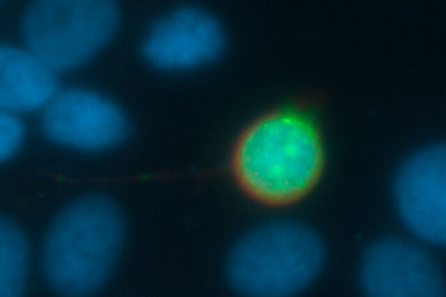
Iron metabolism plays a role in cancer development and progression, and modulates the immune response. Understanding how iron influences cancer and the immune system can aid the development of new therapeutic approaches.

Going Beyond Deconvolution

Widefield fluorescence microscopy is often used to visualize structures in life science specimens and obtain useful information. With the use of fluorescent proteins or dyes, discrete specimen components are marked in a highly specific manner. To fully understand a structure, visualizing it in 3 dimensions can be necessary, but certain challenges are faced when doing so with microscopy.

Find Relevant Specimen Details from Overviews
Switch from searching image by image to seeing the full overview of samples quickly and identifying the important specimen details instantly with confocal microscopy. Use that knowledge to set up high-resolution image acquisition automatically using templates for slides, dishes, and multi-well plates. Like a GPS for the cells of a specimen, users always have a clear path to high-quality data with the LAS X Navigator software, a powerful navigation tool for the STELLARIS platform. LAS X Navigator enables confocal and fluorescence lifetime experiments to be combined with a stage application and makes all the TauSense tools available for use.

How to Remove Out-Of-Focus Blur and Improve Segmentation Accuracy
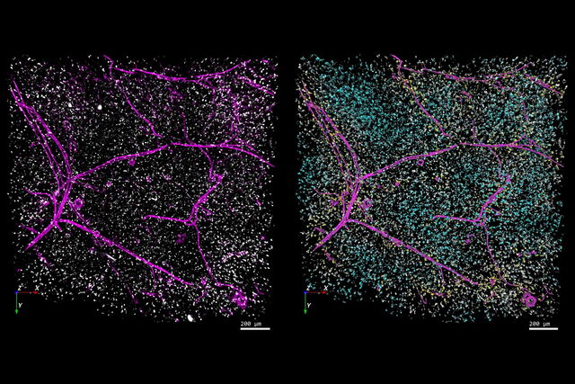
The specificity of fluorescence microscopy allows researchers to accurately observe and analyze biological processes and structures quickly and easily, even when using thick or large samples. However, out-of-focus fluorescence increases background, reduces contrast, and makes accurate image segmentation more challenging. A powerful solution to these issues is the use of THUNDER to remove image blur in combination with Aivia which automates widefield image analysis using AI to improve speed and accuracy. Here, we explore this synergetic approach in more detail.

THUNDER Imaging Systems

Easily tackle biologically relevant 3D models with THUNDER Imagers.

They bring you high-speed, multicolor imaging of thin and thick samples with increased temporal resolution in the first attempt itself.

To answer important scientific questions, they enable you to obtain a clear view of details, even deep within an intact sample, in real time without out-of-focus blur. Sharp imaging of 2D and 3D specimens is now as easy as working with your favorite camera-based fluorescence microscope.
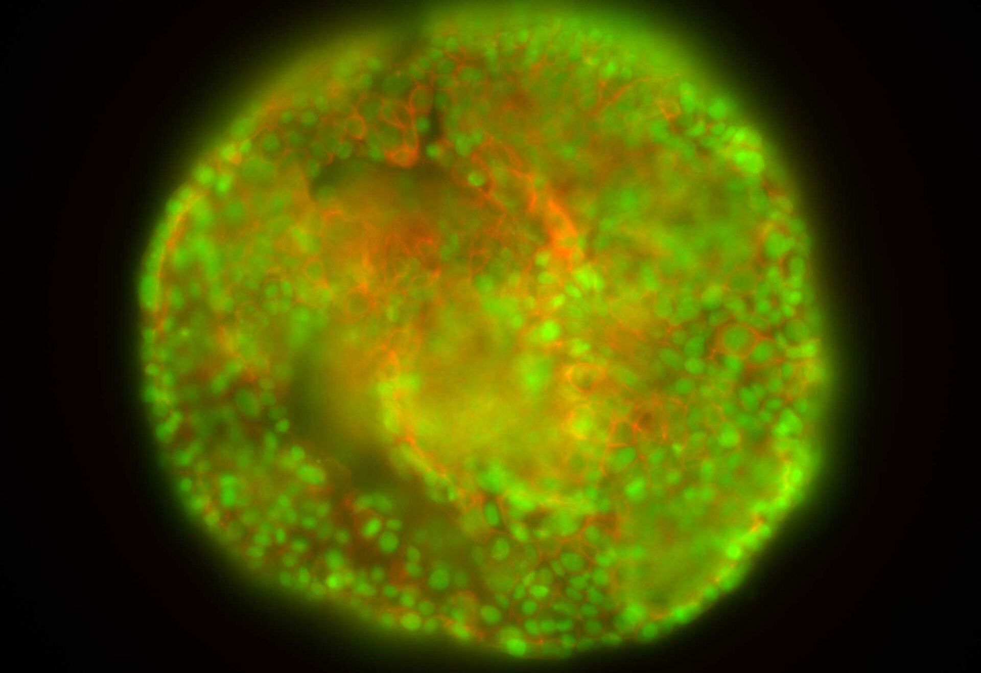
Standard FluorescenceTHUNDER Imager

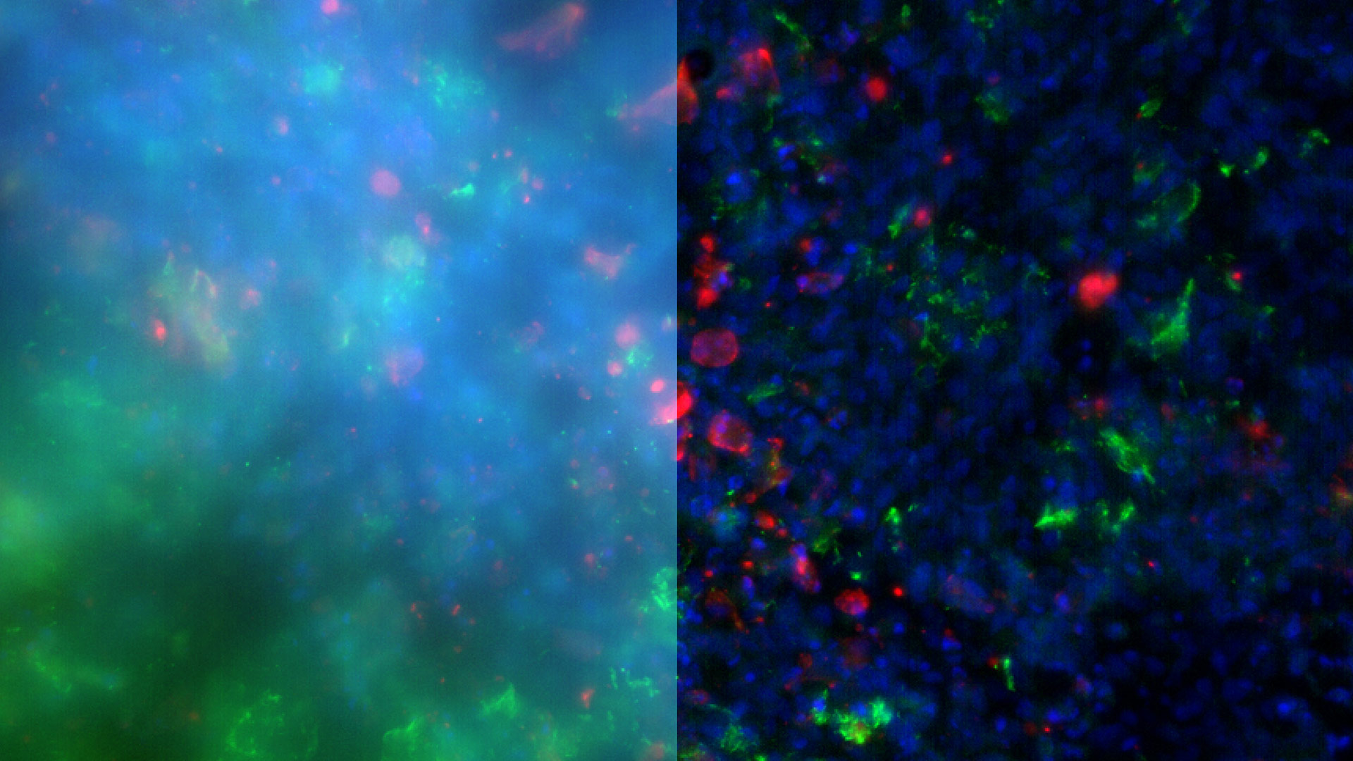
HeLa cell spheroid stained with Alexa Fluor 568 Phalloidin (Actin) and YOYO 1 iodide (Nucleus).

Decode 3D biology in real time*

Fundamentally change the way you work when imaging model organisms, tissue sections, and 3D cell cultures like organoids. Whether single cells, tissues, whole organisms, or tumor spheroids, THUNDER Imagers enable the decoding of 3D biology in real time, with immediate visual feedback.

THUNDER imaging systems excel due to

  • Delivery of benchmark performance and first-rate results for your application
  • Clear view of details even deep within the specimen thanks to Computational Clearing, directly in your live preview
  • Ease of use, speed, and sensitivity, just like with widefield imaging


*in accordance with ISO/IEC 2382:2015

image analysis software solutions

Analyze your images for hidden insights

Aivia and LAS X, advanced image analysis software solutions from Leica Microsystems empower you to take your microscopy-based research to the next level.

By combining THUNDER with Leica’s AI-driven image analysis software Aivia, you can gain meaningful insights from your microscopy imaging data through a wide range of machine learning and deep learning algorithms. Learn more about image analysis solutions from Leica Microsystems.
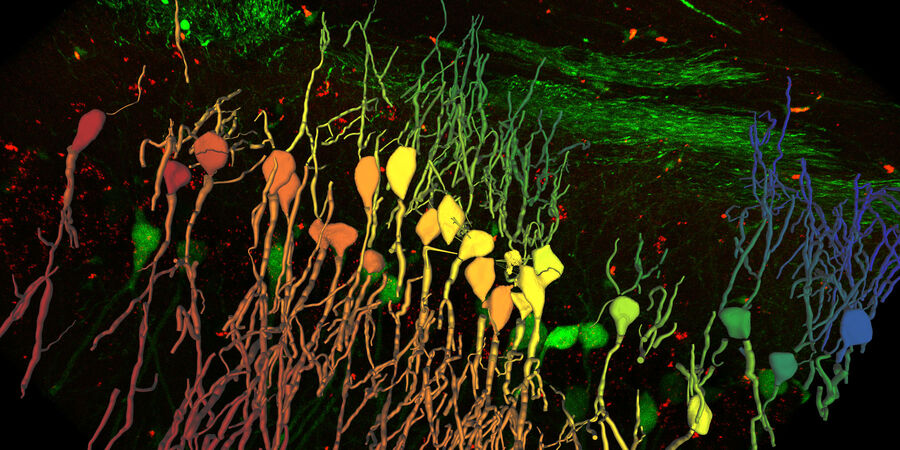
image analysis solutions

Take a look at all our upcoming congresses, exhibitions, webinars, and workshops and join us at one of our next events!

All events

14
19 Sep 2026

IMTS 2026

United States Tradeshow

The THUNDER Technology

THUNDER is an opto-digital technology that uses the Computational Clearing method to generate high resolution and high contrast images. Computational Clearing removes the typical haze inherent to all widefield images of thick samples. It produces brilliant results for large image stacks, as well as single images taken deep in your sample.

THUNDER, a Leica technology, automatically takes all relevant optical parameters into account in order to achieve haze-free results in real time.

Technology Note

Still questions about the THUNDER technology? The detailed answers are in this technology note.
Standard FluorescenceTHUNDER Imager

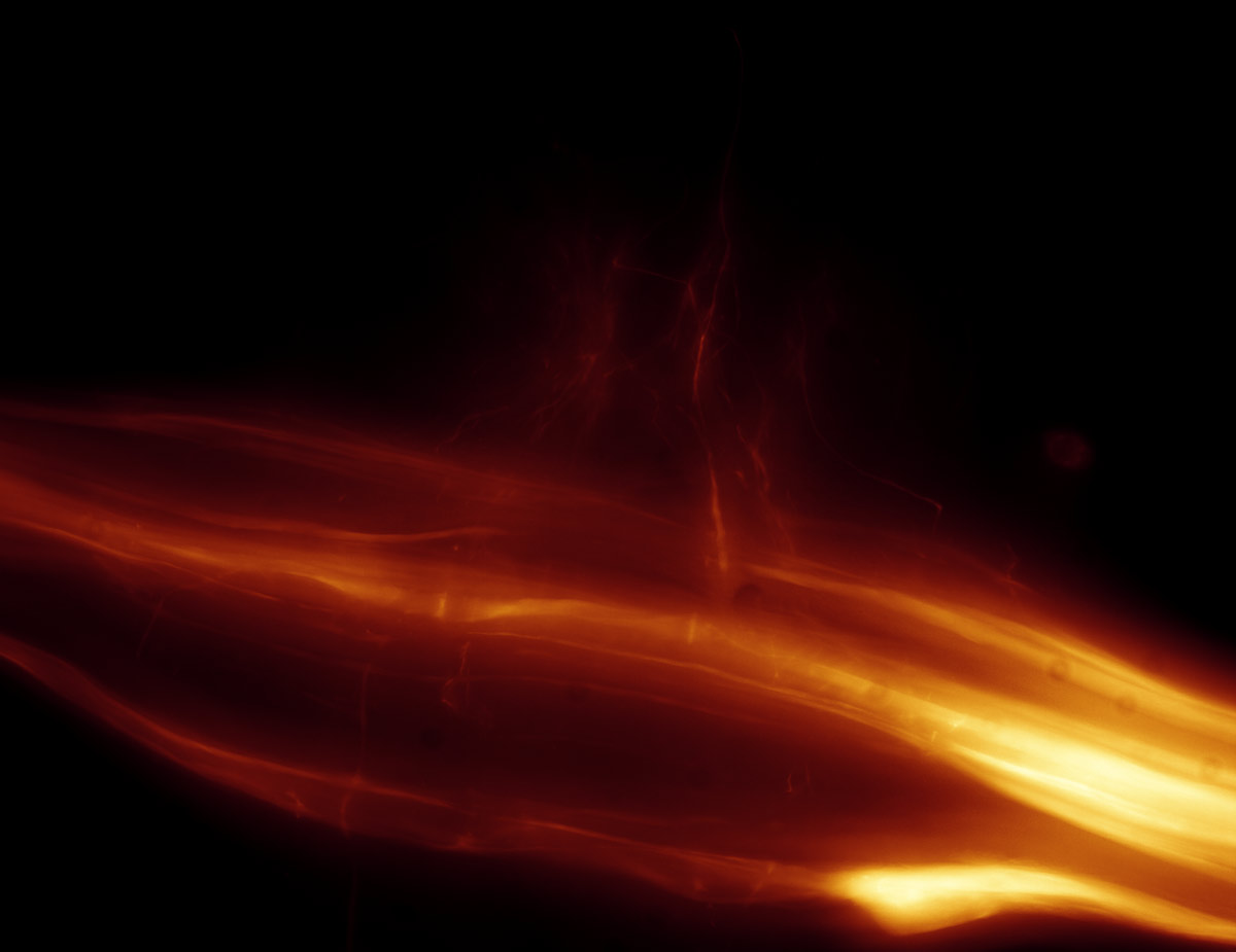
Image of a locust ganglion, showing a maximum projection, without (left) and with (right) THUNDER. Sample thickness: 110µm, data volume: 376 MB. Acquisition time using Computational Clearing: 3 seconds.

Speed you can see - THUNDER Live

Speed You Can See

Show details

Speed You Can See

Perform high resolution tissue imaging as well as live sample and model organism imaging on thick samples without struggling to find your region of interest.

The new THUNDER Live add-on visualizes a computationally cleared image instantly in the live viewer and allows you to optimize ICC parameters by using live image feedback.

THUNDER offers speed that you can see and feel.

Imaging a mouse brain section

Show details

Imaging a mouse brain section with THUNDER Live

Focusing through a 165µm thick mouse brain section using THUNDER Live. Feature scale is used to emphasize neurites. Neurites are much better visualized with THUNDER Live compared to conventional widefield microscopy, allowing better identification of regions of interest. In addition, THUNDER Live shows a preview of THUNDER image quality.

Neurons expressing Thy1-YFP fusion protein. Objective: 63x/1.4 oil.

Sample courtesy of Dr. Hong Xu, University of Pennsylvania, Philadelphia (USA)

Imaging a tumor spheroid

Show details

Imaging a tumor spheroid with THUNDER Live

With THUNDER Live, it is possible to evaluate the dynamics of cell-cell and cell-matrix interactions in deeper region of a 3D cell culture sample in real time. Without THUNDER Live, a user using conventional widefield microscopy, would struggle to see intrinsic details in depth due to out of focus blur. Now with THUNDER Live, the user can unlock details that were otherwise masked by the out of focus blur in depth and determine suitability of regions of interest for imaging or creating 3D volumes with live preview. 

Imaging a mouse kidney section

Show details

Imaging a mouse kidney section with THUNDER Live

Focusing through a 20µm thick mouse kidney section. Feature scale was used to visualize proximal tubule boarders allowing better identification of regions of interest compared to conventional widefield microscopy.

Achieve Outstanding Results with Ease

THUNDER works fast. All it takes is one click. No need to calibrate the system or to adjust hardware components. Just select a Computational Clearing method and generate expert-level results in a breathtakingly short time.

Need to replicate an experiment for multiple specimens? All the imaging parameters of THUNDER can be easily reproduced and applied to other tasks.
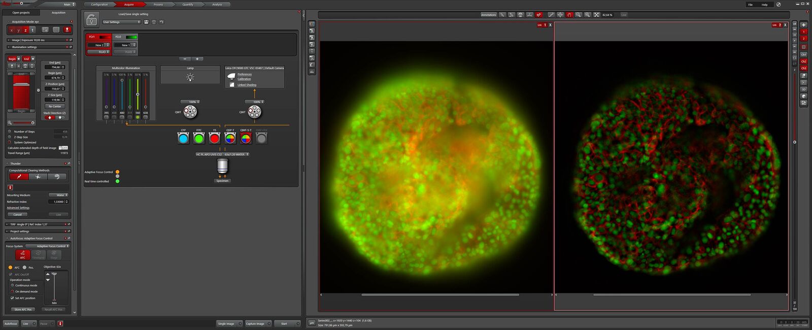
THUNDER would specially be useful for time-lapse, because it allows very fast scanning of big samples in less than 2 minutes, and provides exceptionally crisp images.

Dr. Almary Guerra, Max Planck Institute for Heart and Lung Research, Bad Nauheim (Germany)

Computational Clearing

Computational Clearing efficiently differentiates between wanted signal and background by taking the feature size of a specimen into account. This approach instantly makes image details visible which may have been formerly obscured by unwanted signal. With the need of only a single image, stunning results can be displayed live for the user to view. It provides a powerful technique for both 2D and 3D imaging.

Depending on the type of application, computational clearing can be combined with adaptive deconvolution using the Leica decision mask technology. With the method being fully automated, it can work independent of manual user input. THUNDER delivers consistently high-quality images, surpassing standard widefield images. Improving multi-dimensional data is only a single click away.

Reliable results, ready for the next step

Just activate Computational Clearing once and acquire haze-free images. It does not matter how simple or complex your experiments are, the raw data will always be stored for validation.

The haze free results with clear structures can easily be segmented and used for further analysis, such as counting nuclei, or the number of spots per nucleus, particle tracking, etc.

See through the haze

THUNDER Imagers remove the out-of-focus blur that occurs with widefield observation through the Leica method called Computational Clearing. With THUNDER Imagers you can have both high-quality 3D images of thick samples and, at the same time, benefit from the speed and sensitivity like with a widefield system.
Zebrafish Research

For the best result during screening, sorting, manipulation, and imaging you need to see details and structures to make the right decisions for your next steps in research. Known for outstanding optics and superb resolution, stereo microscopes and transmitted light bases from Leica are the preferred choice of researchers worldwide. 

Read More

Life Science Research

Leica Microsystems’ life science research microscopes support the imaging needs of the scientific community with advanced innovation and technical expertise for the visualization, measurement and analysis of microstructures.

Read More

Live Cell Imaging

Shifting perspective from single microscope components to a full working live cell imaging solution, Leica Microsystems integrates microscope, LAS X imaging software, cameras, and dedicated third-party components into a complete live cell imaging system.

Read More

Fluorescence Microscopy

Fluorescence is one of the most commonly used physical phenomena in biological and analytical microscopy, mainly because of its high sensitivity and high specificity. Find out how fluorescence microscopes support your research.

Read More

Zebrafish Research

For the best result during screening, sorting, manipulation, and imaging you need to see details and structures to make the right decisions for your next steps in research. Known for outstanding optics and superb resolution, stereo microscopes and transmitted light bases from Leica are the preferred choice of researchers worldwide. 

Read More

Life Science Research

Leica Microsystems’ life science research microscopes support the imaging needs of the scientific community with advanced innovation and technical expertise for the visualization, measurement and analysis of microstructures.

Read More

Scroll to top