Contact Us

Leica Microsystems

Leica Microsystems

Leica Microsystems is a world leader in microscopes and scientific instruments. Founded as a family business in the nineteenth century, the company’s history was marked by unparalleled innovation on its way to becoming a global enterprise.

Its historically close cooperation with the scientific community is the key to Leica Microsystems’ tradition of innovation, which draws on users’ ideas and creates solutions tailored to their requirements. At the global level, Leica Microsystems is organized in three divisions, all of which are among the leaders in their respective fields: Life Science, Industry and Medical.

The company is represented in over 100 countries with 6 manufacturing facilities in 5 countries, sales and service organizations in 20 countries, and an international network of dealers. The company is headquartered in Wetzlar, Germany.
http://www.leica-microsystems.com/

Primary Beam Splitting Devices for Confocal Microscopes

Current fluorescence microscopy employs incident illumination which requires separation of illumination and emission light. The classical device performing this separation is a color-dependent beam…
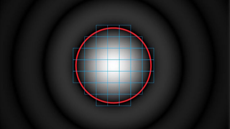
Pinhole diameter and diffraction pattern.

Pinhole Effect in Confocal Microscopes

When operating a confocal microscope, or when discussing features and parameters of such a device, we inescapably mention the pinhole and its diameter. This short introductory document is meant to…
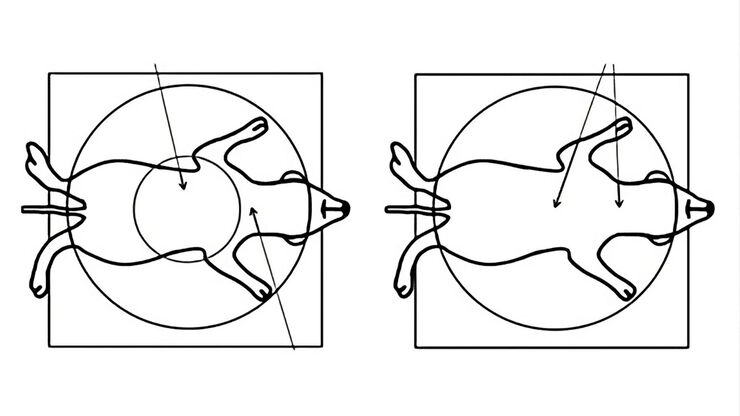
Comparison when observing a rodent model organism with a Greenough versus CMO (common main objective) stereo microscope for a task like surgery.

Rodent and Small-Animal Surgery

Learn how you can perform rodent (mouse, rat, hamster) and small-animal surgery efficiently with a microscope for developmental biology and medical research applications by reading this article.
Image Guided Surgery

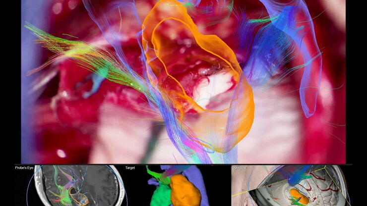
Navigating Through the Brain

One of the challenges of neurosurgery is orientation at the surgical site. When resecting tumors, removing arteriovenous malformations, or clipping aneurysms, surgeons often have to work near healthy…
Left: Tissue cells marked with an immunolabel (FITC) illuminated with wide-band UV excitation. Note the tissue structure with blue autofluorescence. Right: Same tissue and same immunostaining with FITC label illuminated with epi-illumination using narrow-band blue (490 nm) light. Note the increased image contrast (Ploem, 1967)

Milestones in Incident Light Fluorescence Microscopy

Since the middle of the last century, fluorescence microscopy developed into a bio scientific tool with one of the biggest impacts on our understanding of life. Watching cells and proteins with the…

Porous Ceramics - Sample Preparation for SEM

Application Note for Leica EM RES102 - Ceramic membrane filters with pore sizes down to a few nanometres must be investigated in cross-section with regard to the structure of the pores. The smallest…
Coaxial light with only polarizer open.

Digital Microscopy with Versatile Illumination and Various Contrast Methods for More Efficient Inspection and Quality Control

State-of-the-art digital microscopes utilizing a versatile illumination system capable of achieving multiple contrast methods, such as the Leica DVM6, are very useful for inspection, quality control,…
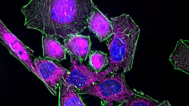
HeLa cells stimulated with LPS. Image has been subjected to deconvolution.

Chronic Inflammation Under the Microscope

In the course of chronic inflammation certain body areas are recurrently inflamed. This goes along with many human diseases. With the help of widefield light microscopy, the underlying processes can…

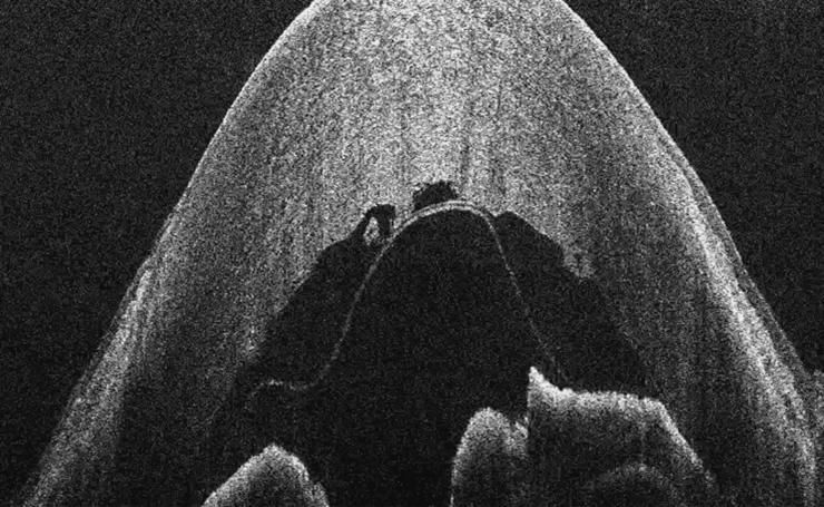
What is OCT?

Optical Coherence Tomography (OCT) is a non-invasive, non-contact imaging modality used to visualize and monitor changes to the morphology of biological tissue. OCT employs low-coherence…
Wifi education solutions

Factors for Selecting Student Microscopes

If chosen carefully, educational microscopes will open windows to a cosmos of minute detail which delight young minds in schools and universities – and ideally keeps them fascinated enough to make…
Gene editing with Cas9

Gene Editing with CRISPR/Cas9 - Breakthrough in Genome Engineering

The CRISPR/Cas9 system is one of several different bacterial systems for defense against viral attacks. It consists of two main components. One is a small piece of RNA which binds to the viral target…

Paper Samples - Sample Preparation for SEM

Application Note for Leica EM RES102 - A coated paper sample has been prepared with ion beam slope cutting in order to test the procedure with regard to its applicability. With the use of ion beam…
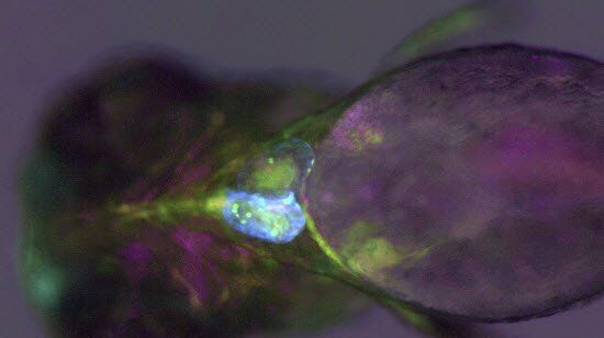
Transgenic zebrafish larva where fluorescent proteins label the heart muscle blue, blood and blood vessels red, and all circulatory system cells green. Image recorded with a M205 FA microscope.

Imaging and Analyzing Zebrafish, Medaka, and Xenopus

Discover how to image and analyze zebrafish, medaka, and Xenopus frog model organisms efficiently with a microscope for developmental biology applications from this article.

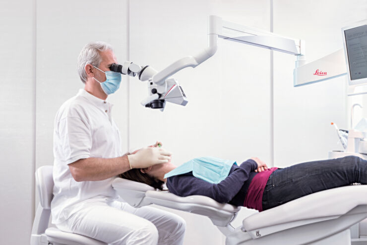
Successful Endodontic Treatment with Dental Operating Microscopes

In endodontics, accurate treatment is not only dependent on the technical skills and knowledge of the dentist, but also on clear, detailed visualization of the surgical field. As the outcome of an…
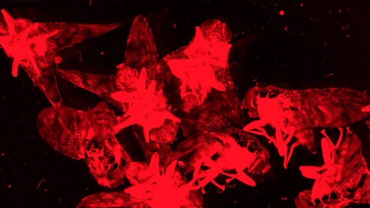
Fluorescence stereo microscope image of anesthetized Mediterranean fruit flies recorded with a M205 stereo microscope.

Investigating Fruit Flies (Drosophila melanogaster)

Learn how to image and investigate Drosophila fruit fly model organisms efficiently with a microscope for developmental biology applications from this article.
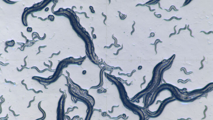
C. elegans

Studying Caenorhabditis elegans (C. elegans)

Find out how you can image and study C. elegans roundworm model organisms efficiently with a microscope for developmental biology applications from this article.

Ion Beam Polishing of Sample Surfaces - Sample Preparation for SEM

Application Note for Leica EM RES102 - Ion milling can be used to reduce the roughness of sample surfaces. Small angles less than 6° with respect to the sample surface are necessary. The high voltage…

Six Features to Consider when Choosing a Dental Microscope

In dental medicine, the surgical microscope has become increasingly important for high-quality and successful surgeries, particularly in the field of endodontics. A microscope supports the dentist to…
Scroll to top