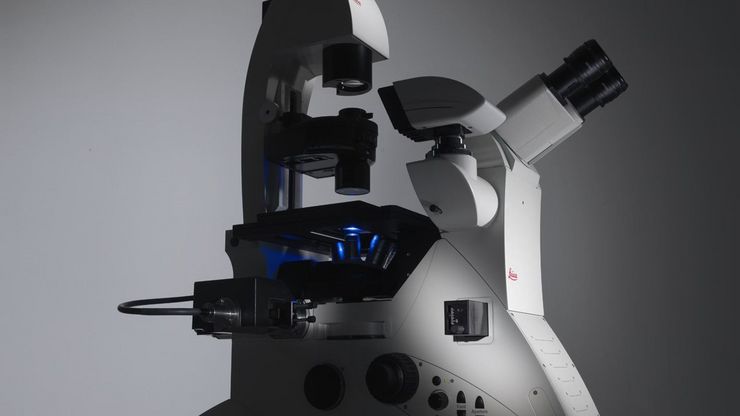
Leica LMD Systems Laser Microdissection Microscopes

Dissection Perfection

Read our latest articles
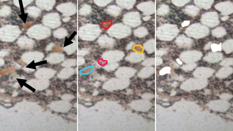
Low-input spatial proteomics workflow for megakaryocyte isolation from bone marrow.

Spatial Proteomics Workflow in Blood Cancer (MPNs)
Megakaryocytes play a central role in the biology of myeloproliferative neoplasms (MPNs), yet their in vivo proteomic characterization remains a major challenge due to low abundance and disrupted…
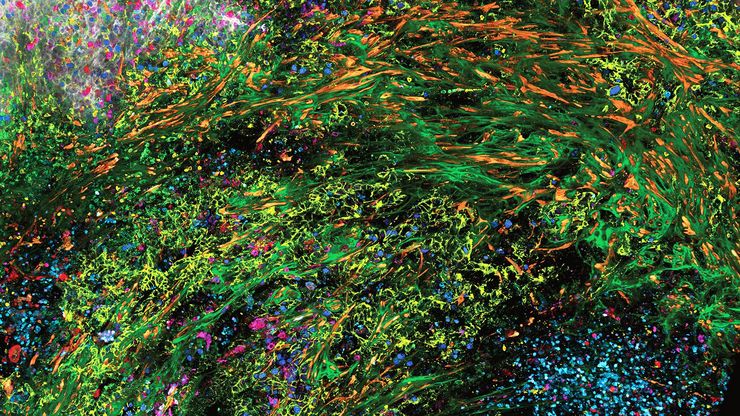
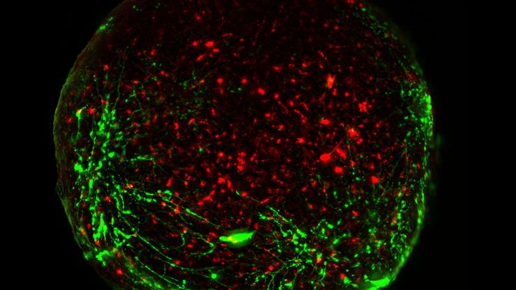
Virally labeled neurons (red) and astrocytes (green) in a cortical spheroid derived from human induced pluripotent stem cells. THUNDER Model Organism Imager with a 2x, 0.15 NA objective at 3.4x zoom was used to produce this 425 µm Z-stack (26 positions) which is presented here as an Extended Depth of Field (EDoF) projection. Images courtesy of Dr. F. Birey, Dr. S. Pasca laboratory, Palo Alto, CA.

Guide to Live-Cell Imaging

For a wide range of applications in various research fields of life science, live-cell imaging is an indispensable tool for visualizing cells in a state as close to in vivo, i.e. living and active, as…
Shown is the DMi8 inverted microscope which is used for life-science research.

Factors to Consider When Selecting a Research Microscope
An optical microscope is often one of the central devices in a life-science research lab. It can be used for various applications which shed light on many scientific questions. Thereby the…
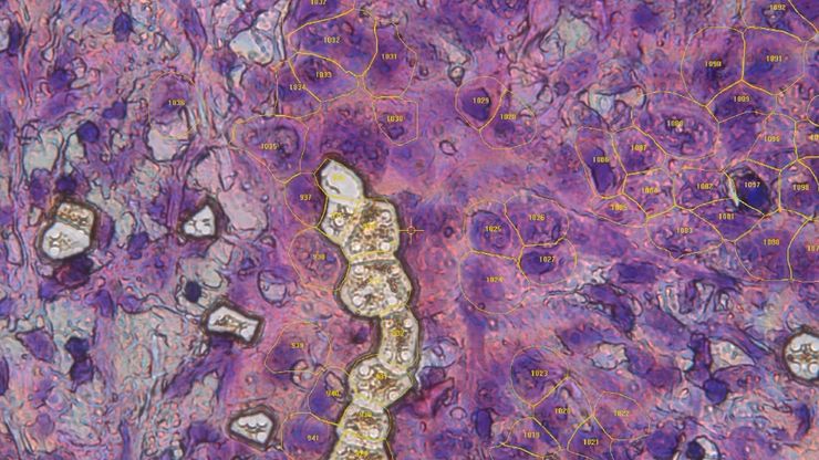
Single cells collected via laser microdissection as part of the Deep Visual Proteomics workflow.

AI meets Deep Visual Proteomics (DVP) to Advance Disease Research
In this webinar, Dr. Andreas Mund will introduce a cutting-edge platform that merges Deep Visual Proteomics (DVP) with AI-powered pathology models, enabling high-resolution mapping of key regions in…
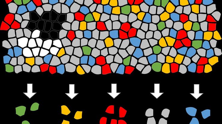
Artificial Intelligence (AI) segmentation used in conjunction with LMD to increase discovery throughput.

Biomarker Discovery with Laser Microdissection

Explore the potential of spatial proteomics workflows, such as Deep Visual Proteomics (DVP), to decipher pathology mechanisms and uncover druggable targets. Altered protein expression, abundance, or…

How a Breakthrough in Spatial Proteomics Saved Lives
Toxic epidermal necrolysis (TEN) is a rare but devastating reaction to common medications like antibiotics or gout treatments. It begins innocuously, often as a rash, but can escalate rapidly into…

A Novel Laser-Based Method for Studying Optic Nerve Regeneration

Optic nerve regeneration is a major challenge in neurobiology due to the limited self-repair capacity of the mammalian central nervous system (CNS) and the inconsistency of traditional injury models.…
Mouse brain slice which was immunostained with GFAP-A647 and imaged using a THUNDER Imager Tissue. Courtesy of H. Xu, University of Pennsylvania, Philadelphia, USA.

A Guide to Neuroscience Research

Neuroscience often requires investigating challenging specimens to better understand the nervous system and disorders. Leica microscopes helps neuroscientists obtain insights into neuronal functions.
Pancreatic Ductal Adenocarcinoma with 11 Apoptosis biomarkers shown – BAK, BAX, BCL2, BCLXL, Caspase9, CIAP1, NaKATPase, PCK26, SMAC, Vimentin, and XIAP.

Transforming Research with Spatial Proteomics Workflows
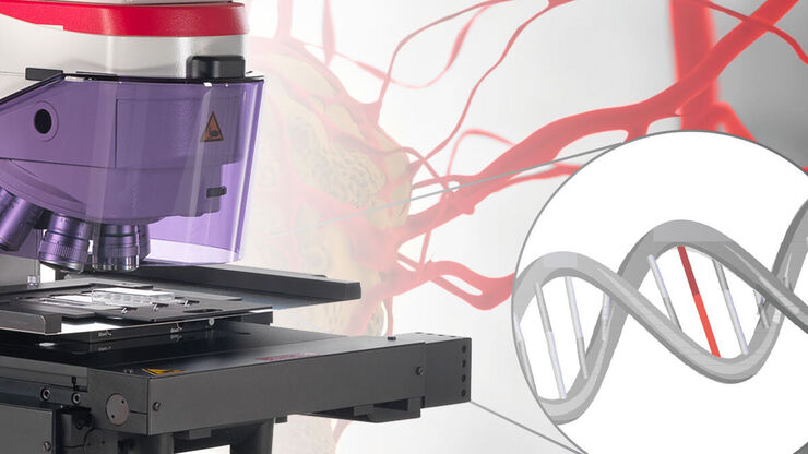
Spatial Proteomics, Nature Methods 2024 Method of the Year, is driving research advancements in cancer, immunology, and beyond. By combining positional data with high throughput imaging of proteins in…
Automated Laser Microdissection for Proteome Analysis

Deep Visual Proteomics Provides Precise Spatial Proteomic Information

Despite the availability of imaging methods and mass spectroscopy for spatial proteomics, a key challenge that remains is correlating images with single-cell resolution to protein-abundance…
Pancreatic Ductal Adenocarcinoma imaged with Cell DIVE. Analysis done by Aivia.

A Guide to Spatial Biology

What is spatial biology, and how can researchers leverage its tools to meet the growing demands of biological questions in the post-omics era? This article provides a brief overview of spatial biology…
Image of a 12-μm thick brain section, which was stained with Toluidin blue, before dissection. It was taken with a microscope using a 63x objective.

An Introduction to Laser Microdissection
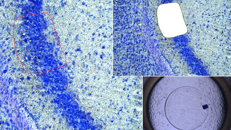
The heterogeneity of histological and biological specimens often requires isolation of specific single cells or cell groups from surrounding tissue before molecular biology analysis can be carried…
Mouse brain (left) microdissected with a 10x objective (upper right). Inspection of the collection device (lower right).

Molecular Biology Analysis facilitated with Laser Microdissection (LMD)
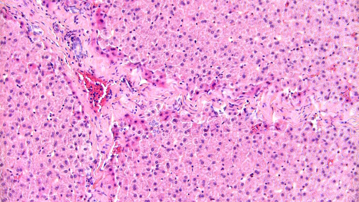
Extracting biomolecules, proteins, nucleic acids, lipids, and chromosomes, as well as extracting and manipulating cells and tissues with laser microdissection (LMD) enables insights to be gained into…
Brightfield image of a pig liver stained with hematoxylin-eosin (HE).

Spatial Metabolomics: Exploring Tumor Complexity and Therapeutic Insights
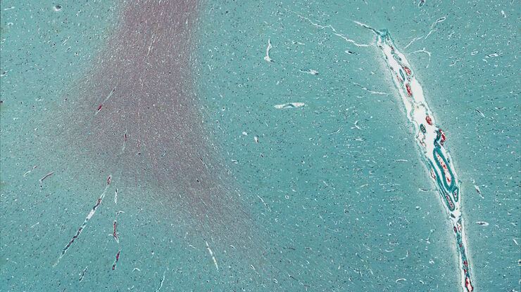
In cancer research, it is vital to understand the interaction between tumor cells and their microenvironment, as the tumor microenvironment influences tumor progression significantly. Spatial…
Mosaic scan of a Masson-Goldner stained cat brain. Magnification: 20x.

Lipidomics Analysis of Sparse Cells based on Laser Microdissection

Delve into cellular intricacies with high-coverage targeted lipidomics analysis of sparse cells. This advanced method, integrating Laser Microdissection (LMD) and Liquid Chromatography-Mass…
Image of murine dopaminergic neurons which have been marked for laser microdissection (LMD).

Neuron Isolation in Spatial Context with Laser Microdissection (LMD)
After Alzheimer’s disease, Parkinson’s is the second most common progressive neurodegenerative disease. Before the first symptoms manifest, up to 70% of dopamine-releasing neurons in the mid-brain…

How did Laser Microdissection enable Pioneering Neuroscience Research?

Dr. Marta Paterlini, a Senior Scientist at the Karolinska Institute, shares her experience of using laser microdissection (LMD) in groundbreaking research into adult human neurogenesis and offers…

Laser Microdissection Protocols for Tissue and Cell Isolation - Download free eBook

In this Bio-protocol Selections, we present a collection of open-access, detailed methods papers using LCM to purify and isolate tissues and cells from plants, mouse embryos, cancer cells, neurons,…
Image of immunofluorescently labelled cells where mitochondria are indicated with red, nuclei with blue, and actin with green.

Studying Virus Replication with Fluorescence Microscopy

The results from research on SARS-CoV-2 virus replication kinetics, adaption capabilities, and cytopathology in Vero E6 cells, done with the help of fluorescence microscopy, are described in this…
Single cell datasets

Exploring Subcellular Spatial Phenotypes with SPARCS

Discover spatially resolved CRISPR screening (SPARCS), a platform for microscopy-based genetic screening for spatial subcellular phenotypes at the human genome scale.
Multi-tissue array with 4 markers shown including DAPI, NaKATPase, PanCk, and Vimentin.

Spatial Biology: Learning the Landscape

Spatial Biology: Understanding the organization and interaction of molecules, cells, and tissues in their native spatial context

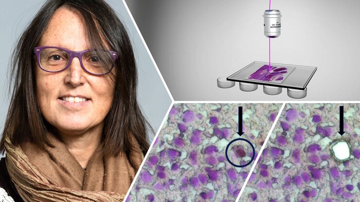
20 Years of Leica Laser Microdissection
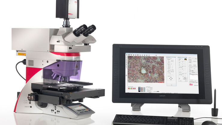
Phenotype-genotype correlations are key for insight. From Eye to Insight is therefore fitting perfectly to Leica Microsystems and in particular to laser microdissection. Laser Microdissection, also…
Leica LMD7

Information about Consumables for Laser Microdissection (LMD)
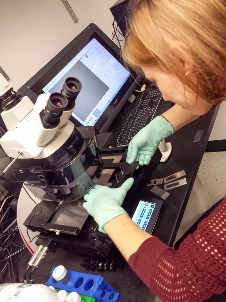
Types of consumables for Leica laser microdissection (LMD) systems, covering from basic to highly specialized applications, are described here. The most common ones are glass membrane slides in…

A Practical Guide to Virology Research
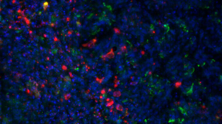
Leica solutions for imaging and sample preparation help you with the investigation of viral entry and fusion, genome integration, viral replication, assembly, and virus budding.

Workflows & Protocols: How to Use a Leica Laser Microdissection System and Qiagen Kits for Successful RNA Analysis

Laser Microdissection (LMD) allows isolating individual cells or chromosomes and is a well established technique for sample preparation prior downstream analysis of the nucleic acid content via PCR or…
SPERM HY-LITER™ staining of a sexual assault smear slide which was performed by a forensic DNA crime laboratory.

Forensic Detection of Sperm from Sexual Assault Evidence
The impact of modern scientific methods on the analysis of crime scene evidence has dramatically changed many forensic sub-specialties. Arguably one of the most dramatic examples is the impact of…
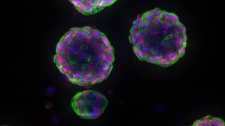
Murine esophageal organoids (DAPI, Integrin26-AF 488, SOX2-AF568) imaged with THUNDER Imager Cell. Courtesy of Dr. F.T. Arroso Martins, Tamere University, Finland.

Biopharma

For biopharma, Leica solutions help accelerate drug discovery, enhance cellular analysis, and support data integrity that meets regulations.
3D high-plex imaging in Cancer Immunology. Detail of a pancreatic tumor section in mouse model, labeled with 15 markers and imaged in one go using STELLARIS SpectraPlex. Total imaged volume 3.2 mm x 2.2 mm x 70 mm in 1 h 20 min. Source: 3D high-multiplex imaging in cancer immunology. Kunz L., Speziale D., et al., Nat. Methods (2024).

Advanced Tissue Imaging & Analysis

Gain insights into tissue structure and function to improve your understanding of spatial biology and disease mechanisms with advanced imaging solutions from Leica Microsystems.

Fields of Application

Biopharma

For biopharma, Leica solutions help accelerate drug discovery, enhance cellular analysis, and support data integrity that meets regulations.

Forensic Science Microscopes

As a forensic scientist, your microscopes and imaging equipment must provide precision, quality, accuracy, and reproducibility of results to ensure success when examining evidence. Leica Microsystems…

Advanced Tissue Imaging & Analysis

Gain insights into tissue structure and function to improve your understanding of spatial biology and disease mechanisms with advanced imaging solutions from Leica Microsystems.

Popular Configurations

Effective April 7, 2025, Leica Microsystems transactions will apply an adjustment tariff charge due to new U.S. tariffs on all applicable products.
EP-DIRECTOR LMD-Slides, 50 slides

EP-DIRECTOR LMD-Slides, 50 slides

Director slides for direct ablation of material with the LMD, pack of 50 slides

Loading...
FLUO FrameSlide, glass-like, 5 pcs

FLUO FrameSlide, glass-like, 5 pcs

Metal frame slides for FLUO, glass like membrane, pack of 5 slides

Loading...
pE 300 lite SB direct fit

pE 300 lite SB direct fit

pE 300 lite SB direct fit

Loading...
PEN Frame Slide(4µm),steel frames,50pcs.

PEN Frame Slide(4µm),steel frames,50pcs.

Metal frame slides with a 4 µm thick PEN membrane, pack of 50

Loading...
PEN Membrane Slide(4µm),glass,50 slides

PEN Membrane Slide(4µm),glass,50 slides

Glass slides with a 4 µm thick PEN membrane, pack of 50

Loading...
PEN MembranSlide, glass, 50 slides

PEN MembranSlide, glass, 50 slides

For laser microdissection special membranes mounted to a glass slide are used to achieve highest quality results. Glass slides covered with a membrane with high UV absorption, making them particularly suitable for cold photo ablation (vaporization), are called glass membrane slides. Applied to the upper surface of the glass slide, the membrane is cemented to the glass at its edges. Thus, a rectangular area to mount the specimen is created where air is between the membrane and the glass backbone. The membrane is specially coated to ensure optimal adhesion of the specimens. In general, glass membrane slides have versatile application potential. Careful treatment of the foils is essential to avoid damage to the membrane. The accumulation of moisture between the glass and the membrane should be avoided, as this can cause the dissectates to stick to the glass backbone.

Loading...
PEN MembranSlide, glass, RNase-free,50pc

PEN MembranSlide, glass, RNase-free,50pc

Glass slides with a 2 µm thick PEN membrane for RNA work, Rnase-free, pack of 50

Loading...
PEN MembranSlides,glass,large,4x25 pcs

PEN MembranSlides,glass,large,4x25 pcs

Large glass slides (50x76 mm) with a 2 µm thick PEN membrane, 4 packs of 25, 100 total slides

Loading...
PEN Petri Dishes 4 µm (1 pack=20 pc)

PEN Petri Dishes 4 µm (1 pack=20 pc)

Petri dishes with 4 µm thick PEN membrane bottom for cutting, pack of 20

Loading...
PEN WhiteCoat(2µm), glass, 200 pcs

PEN WhiteCoat(2µm), glass, 200 pcs

Glass slides with a 2 µm thick PEN membrane, slide has writable area on one end, pack of 200

Loading...
PET FrameSlide, steel frames, 50 slides

PET FrameSlide, steel frames, 50 slides

PET frame slide, a pack of 50 steel frames which can also be used as sample carriers. Leica Microsystems consumables are designed to deliver reliable performance across demanding research workflows. Leica LMD slides and immersion oils work together to support precision, reproducibility, and confidence in laser microdissection and high‑resolution imaging applications. Leica LMD slides are available in multiple membrane types and formats, allowing researchers to select the optimal slide for their sample, detection method, and experimental goal. This flexibility supports both routine workflows and advanced applications such as fluorescence, while demo slide kits make it easy to compare options and identify the best fit.

Loading...
PET FrameSlide,steel frames, RNase-free

PET FrameSlide,steel frames, RNase-free

Metal frame slides with a 1.4 µm thick PET membrane for RNA work, RNase-free, pack of 50

Loading...
POL FrameSlide, steel frames, 50 slides

POL FrameSlide, steel frames, 50 slides

Metal frame slides with a 0.9 µm thick POL membrane, pack of 50

Loading...
POL FrameSlide, steel, 50pc RNase-free

POL FrameSlide, steel, 50pc RNase-free

Metal frame slides with a 0.9 µm thick POL membrane for RNA work, RNase-free, pack of 50

Loading...
PPS FrameSlide (4 µm), steel frames, 50

PPS FrameSlide (4 µm), steel frames, 50

Metal frame slides with a 4 µm thick PPS membrane, pack of 50

Loading...
pre-printed LMD Frame Slides

pre-printed LMD Frame Slides

Metal frame slides with a 4 µm thick PEN membrane with markings for automated shape imports, 10 packs of 5 slides, 50 slides total

Loading...
pre-printed LMD Membrane Slides

pre-printed LMD Membrane Slides

Glass slides with a 4 µm thick PEN membrane with markings for automated shape imports, 10 packs of 5 slides, 50 slides total

Loading...
µ-Slide w. membrane, 18well,ibidi,15 pcs

µ-Slide w. membrane, 18well,ibidi,15 pcs

18-well Ibidi u-slide with 2 µm PEN membrane bottom, pack of 15

Loading...

Don’t see your Configuration? Request for individual Quote

Scroll to top