


機械受容性経路とシナプス経路の研究に顕微鏡がいかに役立つか
このポッドキャストでは、Tobi Langenhan教授は、顕微鏡を使ってシナプスのタンパク質集合体を調べるなど、接着型GPCRの機械受容特性の研究を通して、タンパク質のダイナミクスとその空間的相互作用に精通されています。 Abdullah…

Unlocking Insights in Complex and Dense Neuron Images Guided by AI
The latest advancement in Aivia AI image analysis software provides improved soma detection, additional flexibility in neuron tracing, 3D relational measurement including Sholl analysis and more.
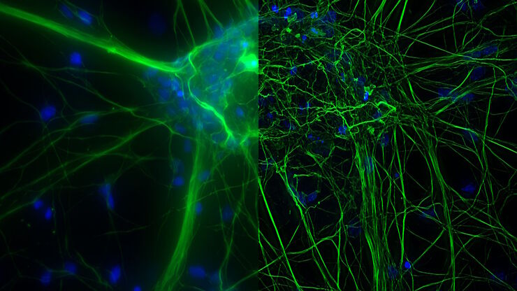
What are the Challenges in Neuroscience Microscopy?
eBook outlining the visualization of the nervous system using different types of microscopy techniques and methods to address questions in neuroscience.
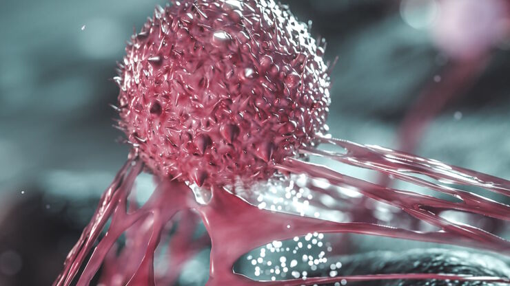
The Role of Iron Metabolism in Cancer Progression
Iron metabolism plays a role in cancer development and progression, and modulates the immune response. Understanding how iron influences cancer and the immune system can aid the development of new…
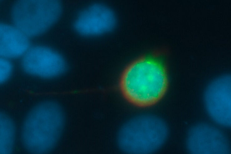
Going Beyond Deconvolution
Widefield fluorescence microscopy is often used to visualize structures in life science specimens and obtain useful information. With the use of fluorescent proteins or dyes, discrete specimen…

3D Spatial Analysis Using Mica's AI-Enabled Microscopy Software
This video offers practical advice on the extraction of publication grade insights from microscopy images. Our special guest Luciano Lucas (Leica Microsystems) will illustrate how Mica’s AI-enabled…
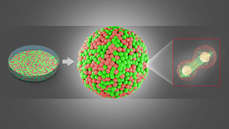
Improve 3D Cell Biology Workflow with Light Sheet Microscopy
Understanding the sub-cellular mechanisms in carcinogenesis is of crucial importance for cancer treatment. Popular cellular models comprise cancer cells grown as monolayers. But this approach…
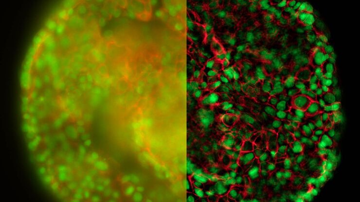
Real Time Images of 3D Specimens with Sharp Contrast Free of Haze
THUNDER Imagers deliver in real time images of 3D specimens with sharp contrast, free of the haze or out-of-focus blur typical of widefield systems. They can even image clearly places deep inside a…

High Resolution Array Tomography with Automated Serial Sectioning
The optimization of high resolution, 3-dimensional (3D), sub-cellular structure analysis with array tomography using an automated serial sectioning solution, achieving a high section density on the…


