Leica Microsystems

Leica Microsystems

Leica Microsystems is a world leader in microscopes and scientific instruments. Founded as a family business in the nineteenth century, the company’s history was marked by unparalleled innovation on its way to becoming a global enterprise.

Its historically close cooperation with the scientific community is the key to Leica Microsystems’ tradition of innovation, which draws on users’ ideas and creates solutions tailored to their requirements. At the global level, Leica Microsystems is organized in three divisions, all of which are among the leaders in their respective fields: Life Science, Industry and Medical.

The company is represented in over 100 countries with 6 manufacturing facilities in 5 countries, sales and service organizations in 20 countries, and an international network of dealers. The company is headquartered in Wetzlar, Germany.
http://www.leica-microsystems.com/

How to make a fast Z-stack

Save time for your 2D and 3D analysis. Watch this video to learn about the new user interface, LAS X.next, for the DVM6 digital microscope. The video demonstrates how to make a fast Z-Stack with a few…

What is a Spectral Detector (SP Detector)?

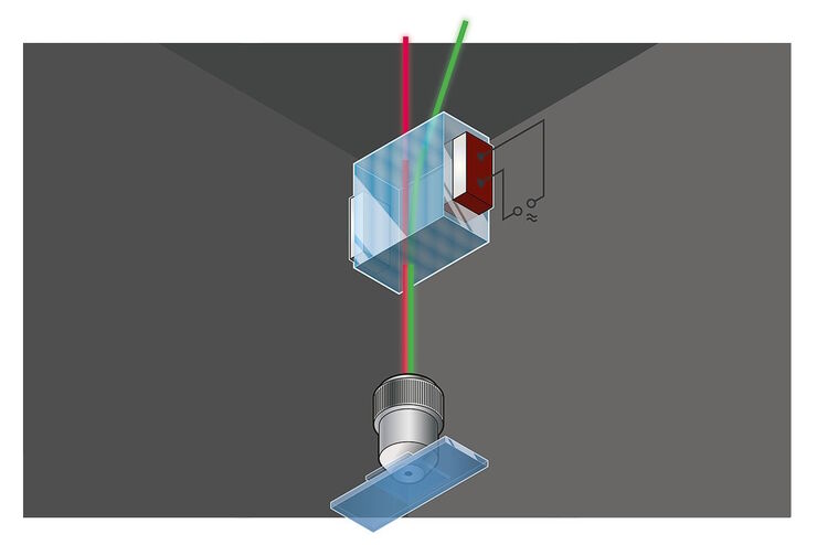
The SP detector from Leica Microsystems denotes a compound detection unit for point scanning microscopes, in particular confocal microscopes. The SP detector splits light into up to 5 spectral bands.…

What is a Resonant Scanner?

A resonant scanner is a type of galvanometric mirror scanner that allows fast image acquisition with single-point scanning microscopes (true confocal and multiphoton laser scanning). High acquisition…
Elucidate cancer development on sub-cellular level by in-vivo like tumor spheroid models.

Improve 3D Cell Biology Workflow with Light Sheet Microscopy

Understanding the sub-cellular mechanisms in carcinogenesis is of crucial importance for cancer treatment. Popular cellular models comprise cancer cells grown as monolayers. But this approach…

What is a Field-of-View Scanner?

A field-of-view scanner is an assembly of galvanometric scanning mirrors used in single-point confocal microscopes that offer the correct optical recording of large field sizes. The field-of-view…

Resolved Field Number (RFN)

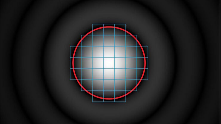
The field number (FN) for optical microscopes indicates the field of view (FOV). It corresponds to the area in the intermediate image that is observable through the eyepieces. Although, we cannot…
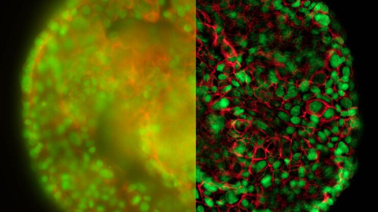
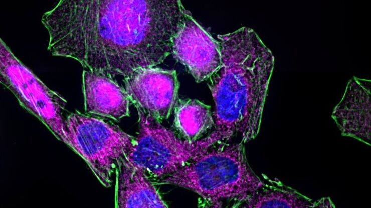
HeLa cell spheroid stained with Alexa Fluor 568 Phalloidin (Actin) and YOYO 1 iodide (Nucleus).

Real Time Images of 3D Specimens with Sharp Contrast Free of Haze

THUNDER Imagers deliver in real time images of 3D specimens with sharp contrast, free of the haze or out-of-focus blur typical of widefield systems. They can even image clearly places deep inside a…

What is a Tandem Scanner?

A Tandem Scanner is an assembly of two different types of scanning together in one system for true confocal point scanning. The Tandem Scanner consists of a three-mirror scanning base with the…
3D glomeruli in a portion of an ECi-cleared kidney scanned by light sheet microscopy. Courtesy of Prof. Norbert Gretz, Medical Faculty Mannheim, University of Heidelberg [1].

Using Mounting Frames for Light Sheet Sample Preparation

Sample handling is an important topic in the context of Light Sheet Microscopy. The TCS SP8 DLS integrates Light Sheet technology into an inverted confocal platform and can hence make use of general…
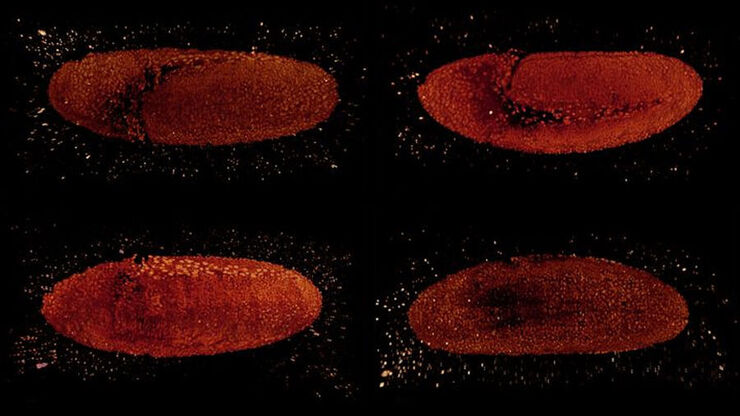
Drosophila embryo observed from different sides. Images were acquired in the framework of the 2018 EMBO Lightsheet Course at MPI CBG in Dresden with expert support of Bruno Cossermelli Vellutini (MPI CBG), Pavel Tomancak (MPI CBG), and Emmanuel Reynaud (UCD).

Using a Rotation Device for Light Sheet Sample Mounting

The TCS SP8 DLS from Leica Microsystems is an innovative concept to integrate the Light Sheet Microscopy technology into the confocal microscope. Due to its unique optical architecture samples can be…

Expanding the Limits of Electron Microscopy Sample Preparation

Capturing the intricate changes in fine structure or in cell dynamics with conventional cryo solutions can be challenging sometimes. Leica Microsystems has developed a new cryo platform, the Leica EM

See More Than Just Your Image

Despite the emergence of new imaging methods in recent years, true 3D resolution is still achieved by Confocal Laser Scanning Microscopy (CLSM). Through a combination of novel, extremely fast scanning…

DIVE Multiphoton Microscope Image Gallery

Today’s life science research focusses on complex biological processes, such as the causes of cancer and other human diseases. A deep look into tissues and living specimens is vital to understanding…

Live Cell Isolation by Laser Microdissection

Laser microdissection is a tool for the isolation of homogenous cell populations from their native niches in tissues to downstream molecular assays. Beside its routine use for fixed tissue sections,…
Array tomography image of T-cells in mouse lymph nodes.

High Resolution Array Tomography with Automated Serial Sectioning

The optimization of high resolution, 3-dimensional (3D), sub-cellular structure analysis with array tomography using an automated serial sectioning solution, achieving a high section density on the…

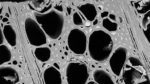
Macroscale to Nanoscale Pore Analysis of Shale and Carbonate Rocks

Physical porosity in rocks, like shale and carbonate, has a large effect on the their storage capacity. The pore geometries also affect their permeability. Imaging the visible pore space provides…

Which Sensor is the Best for Confocal Imaging?

The Hybrid Photodetectors (HyD) are! Why that is the case is explained in this short Science Lab article.

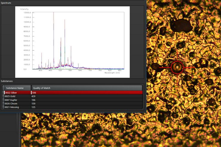
See the Structure with Microscopy - Know the Composition with Laser Spectroscopy

The advantages of a 2-in-1 materials analysis solution combining optical microscopy and laser induced breakdown spectroscopy (LIBS) for simultaneous visual and chemical inspection are described in…

Visualization of DNA Molecules

Precise low angle rotary shadowing with heavy metals (like platinum) can be used in transmission electron microscopy (TEM) to observe molecular details of objects previously absorbed on a thin, low…

The Future of Fluorescence in Vascular Neurosurgery

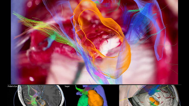
In vascular neurosurgery, surgical microscopes are used to provide a magnified and illuminated view of the surgical field. Although surgeons benefit greatly from the superb image quality and optical…
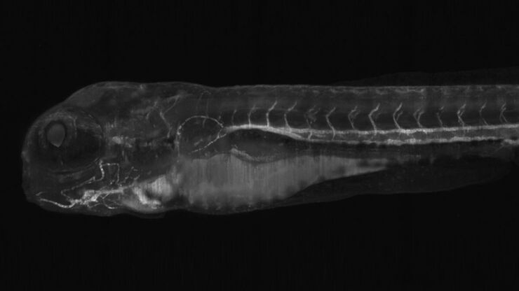
Light sheet microscopy tilescan of zebrafish.

Using U-Shaped Glass Capillaries for Sample Mounting

The DLS microscope system from Leica Microsystems is an innovative concept which integrates the Light Sheet Microscopy technology into the confocal platform. Due to its unique optical architecture,…

Practical Applications of Broad Ion Beam Milling

Mechanical polishing can be time consuming and frustrating. It can also introduce unwanted artifacts when preparing cross-sectioned samples for electron backscatter diffraction (EBSD) in the scanning…
Basalt imaged with crossed polarizers

Digital Microscopy in Earth Science

Classical polarized light (compound) microscopes can only be used for prepared samples, because the working distance they offer is insufficient for whole samples. This means that thicker and bigger…
Multiphoton microscopy of an unstained mouse skin section acquired using the 4Tune detector.

Mission Impossible Accomplished: Tunable Colors for Non-descanning Detection

Leica Microsystems’ 4Tune detector, the key component of the SP8 DIVE Deep In Vivo Explorer, provides spectrally tunable image recording with non-descanning detection. An innovative solution for…
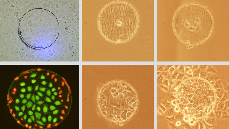
Mammalian cell culure. Phase contrast and fluorescence image.

Introduction to Mammalian Cell Culture

Mammalian cell culture is one of the basic pillars of life sciences. Without the ability to grow cells in the lab, the fast progress in disciplines like cell biology, immunology, or cancer research…

Interview with Dr. Shigeki Watanabe on Research in Synaptic Membrane Dynamics

Dr. Shigeki Watanabe, principle investigator of the department of Cell Biology at the Johns Hopkins University School of Medicine in Baltimore, held a workshop in Zürich, Switzerland on methods to…
„Confetti-Mouse“, taken with the 4Tune spectral detector of Leica’s Deep in-vivo Explorer SP8 DIVE.

Laser Beam Shaping for Multicolor Multiphoton Microscopy

Multiphoton Microscopy is one of the current hot topics in life science research. The new Leica TCS SP8 DIVE from Leica Microsystems presents a series of beneficial new innovations, including a freely…
Convalaria

Introduction to Widefield Microscopy

This article gives an introduction to widefield microscopy, one of the most basic and commonly used microscopy techniques. It also shows the basic differences between widefield and confocal…

Optimization of the Interplay of Optical Components for Aberration Free Microscopy

Optical microscopes are used to magnify objects which are otherwise invisible for the human eye. For this purpose high quality optics is necessary to achieve appropriate resolution. However, besides…

Primary Beam Splitting Devices for Confocal Microscopes

Current fluorescence microscopy employs incident illumination which requires separation of illumination and emission light. The classical device performing this separation is a color-dependent beam…
Scroll to top