
Science Lab
Science Lab
Bienvenido al portal de conocimiento de Leica Microsystems. Aquí encontrará investigación científica y material didáctico sobre el tema de la microscopía. El portal ayuda a principiantes, profesionales experimentados y científicos por igual en su trabajo diario y en sus experimentos. Explore tutoriales interactivos y notas de aplicación, descubra los fundamentos de la microscopía, así como las tecnologías de gama alta. Forme parte de la comunidad Science Lab y comparta sus conocimientos.
Filter articles
Tags
Story Type
Products
Loading...
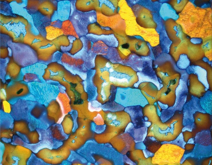
Metallography with Color and Contrast
The examination of microstructure morphology plays a decisive role in materials science and failure analysis. There are many possibilities of visualizing the real structures of materials in the light…
Loading...

Is that Document Genuine or Fake? How do They Identify Fake Documents?
This article shows how forensic experts use microscopy for analysis to identify counterfeit, fake documents, such as ID cards, passports, visas, certificates, etc. Then they know if it is genuine or…
Loading...
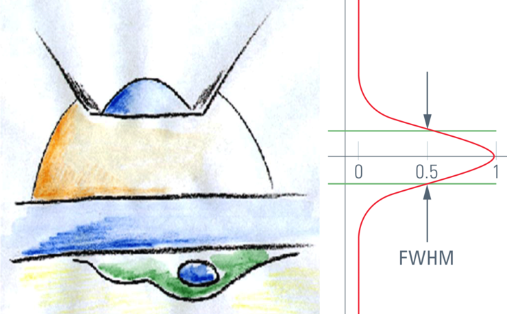
Confocal Optical Section Thickness
Confocal microscopes are employed to optically slice comparably thick samples.
Loading...

Optical Contrast Methods
Optical contrast methods give the potential to easily examine living and colorless specimens. Different microscopic techniques aim to change phase shifts caused by the interaction of light with the…
Loading...

Integrated Modulation Contrast (IMC)
Hoffman modulation contrast has established itself as a standard for the observation of unstained, low-contrast biological specimens. The integration of the modulator in the beam path of themodern…
Loading...

Cochlea Implants for Deaf and Severely Hard of Hearing
The cochlea implant (CI) replaces the function of the outer ear, the middle ear and the cochlea of the inner ear. Basically, it consists of an external unit comprising a microphone, speech processor,…
Loading...
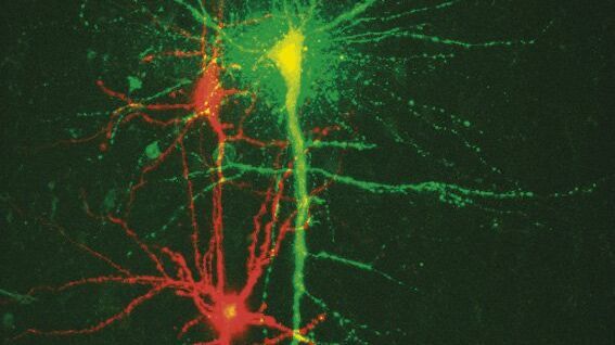
New Standard in Electrophysiology and Deep Tissue Imaging
The function of nerve and muscle cells relies on ionic currents flowing through ion channels. These ion channels play a major role in cell physiology. One way to investigate ion channels is to use…
Loading...

Forensic Detection of Sperm from Sexual Assault Evidence
The impact of modern scientific methods on the analysis of crime scene evidence has dramatically changed many forensic sub-specialties. Arguably one of the most dramatic examples is the impact of…
Loading...

The History of Stereo Microscopy
This article gives an overview on the history of stereo microscopes. The development and evolution from handcrafted instruments (late 16th to mid-18th century) to mass produced ones the last 150…
