
Science Lab
Science Lab
Benvenuti nel portale delle conoscenze di Leica Microsystems. Troverete materiale didattico e di ricerca scientifica sul tema della microscopia. Il portale supporta i principianti, i professionisti esperti e gli scienziati nel loro lavoro quotidiano e negli esperimenti. Esplorate i tutorial interattivi e le note applicative, scoprite le basi della microscopia e le tecnologie di punta. Entrate a far parte della comunità di Science Lab e condividete la vostra esperienza.
Filter articles
Tag
Tipo di storia
Prodotti
Loading...

Intraoperative OCT-Assisted Corneal Transplant Procedures
Learn about the use of intraoperative optical coherence tomography in corneal transplantation and how it facilitates the adaptation of the donor cornea.
Loading...

How Intraoperative OCT Helps Gain Greater Insight in Glaucoma Surgery
Learn about the use of intraoperative Optical Coherence Tomography in glaucoma surgery and how it helps see subsurface tissue details.
Loading...

Ophthalmology: Visualization in Complex Cataract Surgery
Learn about the use of intraoperative Optical Coherence Tomography in cataract surgery and how it supports both standard and complex cataract surgery cases.
Loading...

Improve Macular Hole Surgery with Optical Coherence Tomography
A case study on the use of intraoperative OCT during macular hole surgery for pediatric lamellar macular hole repair and how it provides valuable real-time information.
Loading...

Ophthalmic Gene Therapy Subretinal Injection
Case study on the use of intraoperative OCT for Leber congenital amaurosis macular repair and ophthalmic gene therapy subretinal injection.
Loading...

Dr. Tawfik Shares his Expert View on Direct Horizontal Chopping in Cataract Surgery
It is estimated that nearly 28 million cataract surgery procedures are performed worldwide every year. Phacoemulsification is the most common method used to remove the cataract and chopping maneuvers…
Loading...

How to Select a Microscope for Cataract Surgery
What to consider in the selection of an ophthalmic microscope for cataract procedures. Bearing these aspects in mind will equip surgeons well for talks with manufacturer representatives. Many…
Loading...
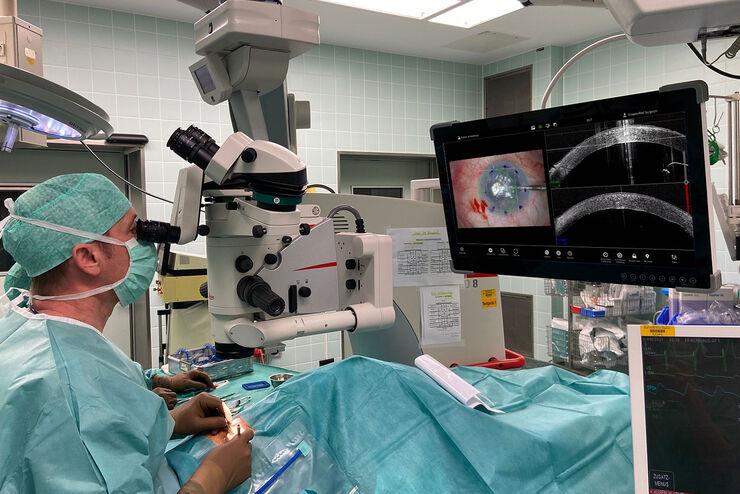
Proveo 8 with intraoperative OCT – a User Evaluation in an University Setting
Optical coherence tomography (OCT) makes structures in the eye visible that lie beneath the surface. When OCT is used intraoperatively, surgeons gain insight into possible pathological changes in the…
Loading...
How to use a Surgical Microscope as an Operating Room Nurse
Surgical microscopes play an essential role in the modern microsurgery procedures. It provides the surgeon, assistant and operating room staff with a magnified and illuminated high-quality image of…
