
Medical Specialties
Medical Specialties
Explore a comprehensive collection of scientific and clinical resources tailored for HCPs, including peer insights, clinical case studies, and symposia. Designed for neurosurgeons, ophthalmologists, and specialists in Plastic and Reconstructive surgery, ENT, and dentistry. This collection highlights the latest advancements in surgical microscopy. Discover how cutting-edge surgical technologies, such as AR fluorescence, 3D visualization, and intraoperative OCT imaging, empower confident decision-making and precision in complex surgeries.

Methods to Improve Reproducibility in Spatial Biology Research
Establish reproducibility results for a Cell DIVE multiplexed imaging study in cancer research using the BAB 200 automated system from ASLS and validated antibodies from CST
 tissue on a single slide.")
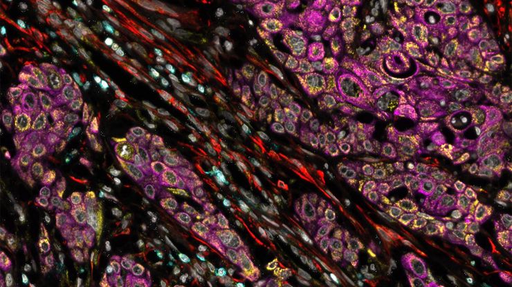
Characterizing tumor environment to reveal insights and spatial resolution
Antibodies from Cell Signaling Technology are validated for use with the Cell DIVE multiplexing workflow and used to probe cell lineages in the tumor microenvironment

Dig Deeper Into the Complexities of Pancreatic Cancer with Multiplex Imaging
Cell DIVE is an iterative staining workflow for multiplexed imaging that unveils biological pathways to dig deeper into the complexities of pancreatic cancer.

How is Microscopy Used in Spatial Biology? A Microscopy Guide
Different spatial biology methods in microscopy, such as multiplex imaging, are helping to better understand tissue landscapes. Learn more in this microscopy guide.

Complex Made Simple: Antibodies in Multiplexed Imaging
Build panels, plan studies, and get the most from precious reagents using this antibody multiplexing guide from Leica Microsystems
Cell DIVE: Unraveling Pathways in Pancreatic Cancer
Unlock new insights into tumour-immune interactions with spatial biology. Join our webinar to learn how to label >60 biomarkers per tissue section and use advanced clustering and analysis techniques…

Confocal Imaging of Immune Cells in Tissue Samples
In this webinar, you will discover how to perform 10-color acquisition using a confocal microscope. The challenges of imaged-based approaches to identify skin immune cells. A new pipeline to assess…

FluoSync - a Fast & Gentle Method for Unmixing Multicolor Images
In this white paper, we focus on a fast and reliable method for obtaining high-quality multiplex images in fluorescence microscopy. FluoSync combines an existing method for hybrid unmixing with…

Multiplexing through Spectral Separation of 11 Colors
Fluorescence microscopy is a fundamental tool for life science research that has evolved and matured together with the development of multicolor labeling strategies in cells tissues and model…
