Industrie
Industrie
Tauchen Sie ein in detaillierte Artikel und Webinare, die sich mit effizienter Inspektion, optimierten Arbeitsabläufen und ergonomischem Komfort in industriellen und pathologischen Umgebungen befassen. Zu den behandelten Themen gehören Qualitätskontrolle, Materialanalyse, Mikroskopie in der Pathologie und vieles mehr. Sie erhalten wertvolle Einblicke in den Einsatz von Spitzentechnologien zur Verbesserung der Präzision und Effizienz von Fertigungsprozessen sowie zur präzisen pathologischen Diagnose und Forschung.
Effiziente Aufnahme von langen Zeitserien
Bei Experimenten mit Aufnahmen langer Zeitserien von Sphäroiden können sich bestimmte Herausforderungen ergeben. Da die Experimente mehrere Tage dauern können, muss die Probe möglichst lange am Leben…
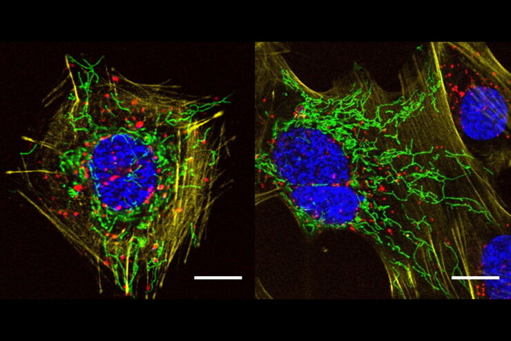
A Versatile Palette of Fluorescent Probes
Researchers at the Max Planck Institute for Medical Research in Heidelberg have developed a general strategy to synthesize live-cell compatible fluorogenic probes, and the result are the new MaP (Max…
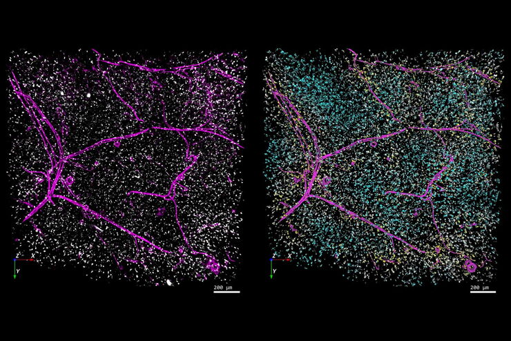
Multicolor 4D Super Resolution Light Sheet Microscopy
The AI Microscopy Symposium offers a unique forum for discussing the latest AI-based technologies and tools in the field of microscopy and biomedical imaging. In this scientific presentation, Yuxuan…
How to Remove Out-Of-Focus Blur and Improve Segmentation Accuracy
The specificity of fluorescence microscopy allows researchers to accurately observe and analyze biological processes and structures quickly and easily, even when using thick or large samples. However,…
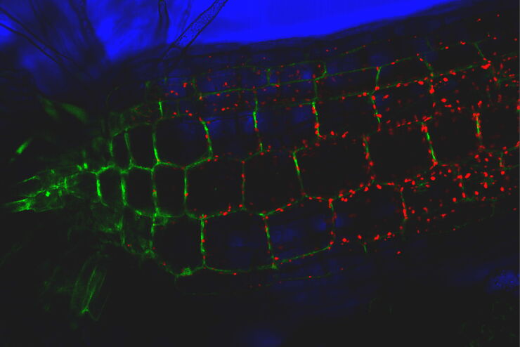
Wie Sie Gewebeproben für die Immunfluoreszenz-Mikroskopie vorbereiten
Immunfluoreszenz (IF) ist eine leistungsfähige Methode zur Visualisierung intrazellulärer Prozesse, Bedingungen und Strukturen. IF-Präparate können mit verschiedenen Mikroskopietechniken (z. B. CLSM,…
Live-Cell Imaging Techniques
The understanding of complex and/or fast cellular dynamics is an important step for exploring biological processes. Therefore, today’s life science research is increasingly focused on dynamic…
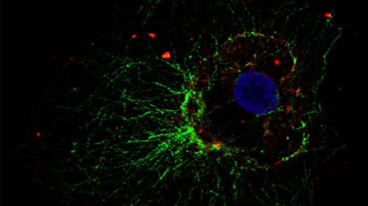
Fluorescent Dyes
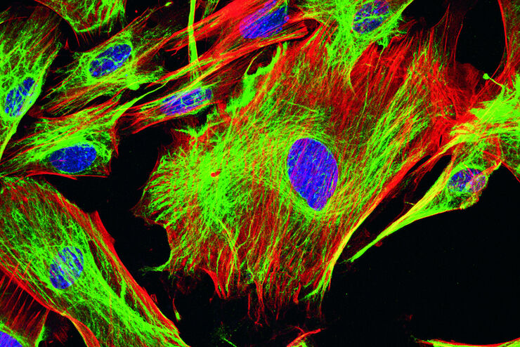
A basic principle in fluorescence microscopy is the highly specific visualization of cellular components with the help of a fluorescent agent. This can be a fluorescent protein – for example GFP –…
The AI-Powered Pixel Classifier
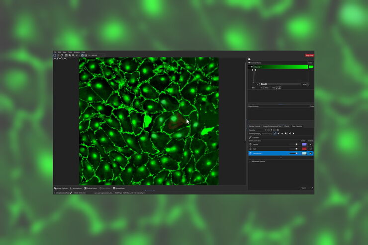
Achieving reproducible results manually requires expertise and is tedious work. But now there is a way to overcome these challenges by speeding up this analysis to extract the real value of the image…
Using Machine Learning in Microscopy Image Analysis
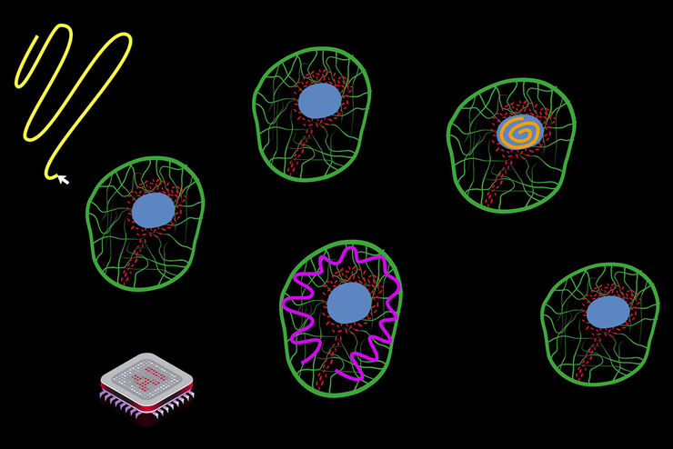
Recent exciting advances in microscopy technologies have led to exponential growth in quality and quantity of image data captured in biomedical research. However, analyzing large and increasingly…

