
Industrie
Industrie
Tauchen Sie ein in detaillierte Artikel und Webinare, die sich mit effizienter Inspektion, optimierten Arbeitsabläufen und ergonomischem Komfort in industriellen und pathologischen Umgebungen befassen. Zu den behandelten Themen gehören Qualitätskontrolle, Materialanalyse, Mikroskopie in der Pathologie und vieles mehr. Sie erhalten wertvolle Einblicke in den Einsatz von Spitzentechnologien zur Verbesserung der Präzision und Effizienz von Fertigungsprozessen sowie zur präzisen pathologischen Diagnose und Forschung.

Regulators of Actin Cytoskeletal Regulation and Cell Migration in Human NK Cells
Dr. Mace will describe new advances in our understanding of the regulation of human NK cell actin cytoskeletal remodeling in cell migration and immune synapse formation derived from confocal and…

Understanding Motor Sequence Generation Across Spatiotemporal Scales
We have developed a microscopy-based pipeline to characterize a developmentally critical behavior at the pupal stage of development, called the ecdysis sequence. We study brain-wide neuronal activity…

Benefits of TauContrast to Image Complex Samples
In this interview, Dr. Timo Zimmermann talks about his experience with the application of TauSense tools and their potential for the investigation of demanding samples such as thick samples or…

Fast, High-quality Vitrification with the EM ICE High Pressure Freezer
The EM ICE High Pressure Freezer was developed with a unique freezing principle and uses only a single pressurization and cooling liquid: liquified nitrogen (LN2). This design enables three major…

Targeting Active Recycling Nuclear Pore Complexes using Cryo Confocal Microscopy
In this article, how cryo light microscopy and, in particular cryo confocal microscopy, is used to improve the reliability of cryo EM workflows is described. The quality of the EM grids and samples is…

Investigating Synapses in Brain Slices with Enhanced Functional Electron Microscopy
A fundamental question of neuroscience is: what is the relationship between structural and functional properties of synapses? Over the last few decades, electrophysiology has shed light on synaptic…

The Power of Pairing Adaptive Deconvolution with Computational Clearing
Learn how deconvolution allows you to overcome losses in image resolution and contrast in widefield fluorescence microscopy due to the wave nature of light and the diffraction of light by optical…
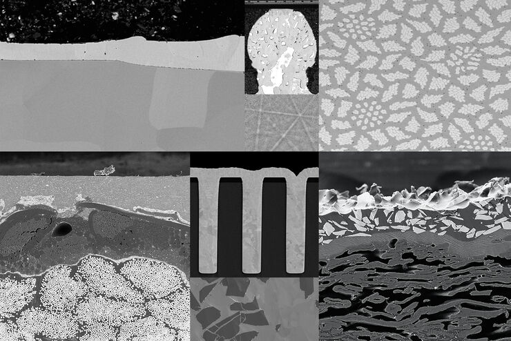
Ion Beam Milling Guide: Enhancing Surface Quality for High-Resolution Imaging and Analysis
In this article you can learn how to optimize the preparation quality of your samples by using the ion beam etching method with the EM TIC 3X ion beam milling machine. A short introduction of the…

Probenvorbereitung für EM: Ein praktischer Leitfaden zur Beschichtung und Gefrierfrakturierung
Von der Niedervakuum-Raumtemperaturbeschichtung bis hin zur Hochvakuum-Kryobeschichtung deckt Leica Microsystems das gesamte Spektrum an Beschichtungsanforderungen ab.
