
Medizinische Fachgebiete
Medizinische Fachgebiete
Entdecken Sie eine umfassende Sammlung wissenschaftlicher und klinischer Ressourcen, die speziell für Ärzte im Gesundheitswesen entwickelt wurden, darunter Berichte von Kollegen, klinische Fallstudien und Symposien. Speziell für Neurochirurgen, Augenärzte, plastische und rekonstruktive Chirurgen, HNO-Ärzte und Zahnärzte. Diese Sammlung präsentiert die neuesten Fortschritte in der chirurgischen Mikroskopie. Entdecken Sie, wie modernste chirurgische Technologien wie AR-Fluoreszenz, 3D-Visualisierung und intraoperative OCT-Bildgebung eine sichere Entscheidungsfindung und Präzision bei komplexen Eingriffen ermöglichen.
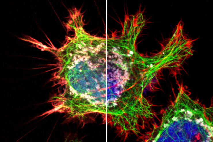
, tubulin with Cy5 (red), and nuclei with DAPI (blue). Image courtesy of Dr. Günter Giese, Max Planck Institute for Medical Research, Heidelberg, Germany.")
Overview of Fluorescent Dyes in terms of Applications and Properties
An introduction to commonly used fluorescent dyes and an overview of their characteristics are given in this article. Fluorescence microscopy is used for the study of specific cellular components with…
, Tropomyosin (cardiomyocytes, red) and GFP (primordial cardiac layer, green).")
A Guide to Fluorescence Microscopy
Fluorescence microscopy uses the ability of fluorophores, dyes, or fluorescent proteins to emit light of a specific wavelength after being excited with light of a shorter wavelength. Biomolecules can…
, actin network (ATTO 647N), and nuclear pore basket (CF 680R).")
The Guide to STED Sample Preparation
This guide is intended to help users optimize sample preparation for stimulated emission depletion (STED) nanoscopy, specifically when using the STED microscope from Leica Microsystems. It gives an…

Epi-Illumination Fluorescence and Reflection-Contrast Microscopy
This article discusses the development of epi-illumination and reflection contrast for fluorescence microscopy concerning life-science applications. Much was done by the Ploem research group…
")
Introduction to Fluorescent Proteins
Overview of fluorescent proteins (FPs) from, red (RFP) to green (GFP) and blue (BFP), with a table showing their relevant spectral characteristics.

An Introduction to Fluorescence
This article gives an introduction to fluorescence and photoluminescence, which includes phosphorescence, explains the basic theory behind them, and how fluorescence is used for microscopy.

Wie Sie Gewebeproben für die Immunfluoreszenz-Mikroskopie vorbereiten
Immunfluoreszenz (IF) ist eine leistungsfähige Methode zur Visualisierung intrazellulärer Prozesse, Bedingungen und Strukturen. IF-Präparate können mit verschiedenen Mikroskopietechniken (z. B. CLSM,…
Explore Innovative Techniques to Separate Fluorophores with Overlapping Spectra
In this article we explore several strategies you can take to improve the separation of fluorophores and increase the number of fluorescent probes you can distinguish in your sample.
The Fundamentals and History of Fluorescence and Quantum Dots
At some point in your research and science career, you will no doubt come across fluorescence microscopy. This ubiquitous technique has transformed the way in which microscopists can image, tag and…
