
Science Lab
Science Lab
Das Wissensportal von Leica Microsystems bietet Ihnen Wissens- und Lehrmaterial zu den Themen der Mikroskopie. Die Inhalte sind so konzipiert, dass sie Einsteiger, erfahrene Praktiker und Wissenschaftler gleichermaßen bei ihrem alltäglichen Vorgehen und Experimenten unterstützen. Entdecken Sie interaktive Tutorials und Anwendungsberichte, erfahren Sie mehr über die Grundlagen der Mikroskopie und High-End-Technologien - werden Sie Teil der Science Lab Community und teilen Sie Ihr Wissen!
Loading...
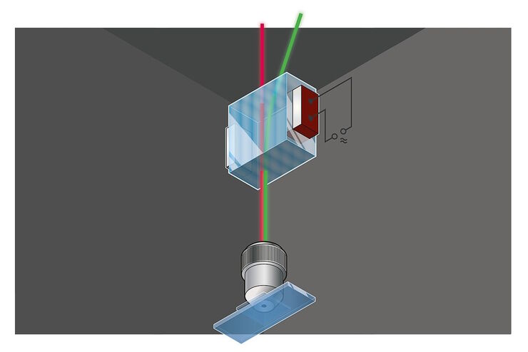
Primary Beam Splitting Devices for Confocal Microscopes
Current fluorescence microscopy employs incident illumination which requires separation of illumination and emission light. The classical device performing this separation is a color-dependent beam…
Loading...

Photoactivatable, Photoconvertible, and Photoswitchable Fluorescent Proteins
Fluorescent proteins (FPs) such as GFP, YFP or DsRed are powerful tools to visualize cellular components in living cells. Nevertheless, there are circumstances when classical FPs reach their limits.…
Loading...

Was macht das Pinhole im konfokalen Mikroskop?
Wer ein konfokales Mikroskop benutzt und über die Eigenschaften und Parameter eines solchen Gerätes diskutiert, wird notwendigerweise über das Pinhole reden – und wie dessen Größe sich auf das…
Loading...
 stereo microscope for a task like surgery.")
Rodent and Small-Animal Surgery
Learn how you can perform rodent (mouse, rat, hamster) and small-animal surgery efficiently with a microscope for developmental biology and medical research applications by reading this article.
Loading...

Navigating Through the Brain
One of the challenges of neurosurgery is orientation at the surgical site. When resecting tumors, removing arteriovenous malformations, or clipping aneurysms, surgeons often have to work near healthy…
Loading...
 illuminated with wide-band UV excitation. Note the tissue structure with blue autofluorescence. Right: Same tissue and same immunostaining with FITC label illuminated with epi-illumination using narrow")
Milestones in Incident Light Fluorescence Microscopy
Since the middle of the last century, fluorescence microscopy developed into a bio scientific tool with one of the biggest impacts on our understanding of life. Watching cells and proteins with the…
Loading...

Video Talk by Kurt Thorn: The Abbe Diffraction Experiment
This lecture describes the famous experiments of Ernst Abbe which showed how diffraction of light by a specimen (and interference with the illuminating light) gives rise to an image and how collection…
Loading...
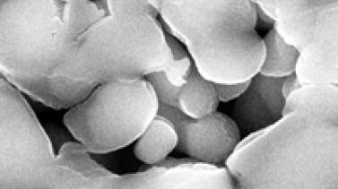
Porous Ceramics - Sample Preparation for SEM
Application Note for Leica EM RES102 - Ceramic membrane filters with pore sizes down to a few nanometres must be investigated in cross-section with regard to the structure of the pores. The smallest…
Loading...

Digital Microscopy with Versatile Illumination and Various Contrast Methods for More Efficient Inspection and Quality Control
State-of-the-art digital microscopes utilizing a versatile illumination system capable of achieving multiple contrast methods, such as the Leica DVM6, are very useful for inspection, quality control,…