
Science Lab
Science Lab
Das Wissensportal von Leica Microsystems bietet Ihnen Wissens- und Lehrmaterial zu den Themen der Mikroskopie. Die Inhalte sind so konzipiert, dass sie Einsteiger, erfahrene Praktiker und Wissenschaftler gleichermaßen bei ihrem alltäglichen Vorgehen und Experimenten unterstützen. Entdecken Sie interaktive Tutorials und Anwendungsberichte, erfahren Sie mehr über die Grundlagen der Mikroskopie und High-End-Technologien - werden Sie Teil der Science Lab Community und teilen Sie Ihr Wissen!
Loading...

Chronic Inflammation Under the Microscope
In the course of chronic inflammation certain body areas are recurrently inflamed. This goes along with many human diseases. With the help of widefield light microscopy, the underlying processes can…
Loading...
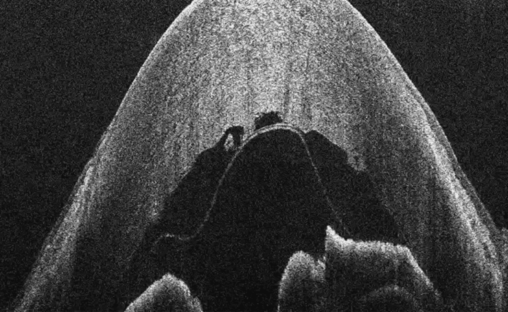
What is OCT?
Optical Coherence Tomography (OCT) is a non-invasive, non-contact imaging modality used to visualize and monitor changes to the morphology of biological tissue. OCT employs low-coherence…
Loading...

Factors for Selecting Student Microscopes
If chosen carefully, educational microscopes will open windows to a cosmos of minute detail which delight young minds in schools and universities – and ideally keeps them fascinated enough to make…
Loading...

Gene Editing with CRISPR/Cas9 - Breakthrough in Genome Engineering
The CRISPR/Cas9 system is one of several different bacterial systems for defense against viral attacks. It consists of two main components. One is a small piece of RNA which binds to the viral target…
Loading...
Paper Samples - Sample Preparation for SEM
Application Note for Leica EM RES102 - A coated paper sample has been prepared with ion beam slope cutting in order to test the procedure with regard to its applicability. With the use of ion beam…
Loading...

Imaging and Analyzing Zebrafish, Medaka, and Xenopus
Discover how to image and analyze zebrafish, medaka, and Xenopus frog model organisms efficiently with a microscope for developmental biology applications from this article.
Loading...
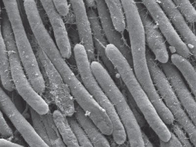
Bacteria Protocol - Critical Point Drying of E. coli for SEM
Application Note for Leica EM CPD300 - Critical point drying of E. coli with subsequent platinum / palladium coating and SEM analysis. Sample was inserted into a filter disc (Pore size: 16 - 40 μm)…
Loading...
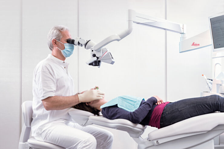
Erfolgreiche endodontische Behandlung mit Dental-Operationsmikroskopen
In der Endodontie hängt die zielgerichtete Behandlung nicht nur von den technischen Fähigkeiten und dem Wissen des Zahnarztes ab. Es kommt auch auf die klare, detaillierte Visualisierung des…
Loading...

Investigating Fruit Flies (Drosophila melanogaster)
Learn how to image and investigate Drosophila fruit fly model organisms efficiently with a microscope for developmental biology applications from this article.