
Science Lab
Science Lab
Bienvenido al portal de conocimiento de Leica Microsystems. Aquí encontrará investigación científica y material didáctico sobre el tema de la microscopía. El portal ayuda a principiantes, profesionales experimentados y científicos por igual en su trabajo diario y en sus experimentos. Explore tutoriales interactivos y notas de aplicación, descubra los fundamentos de la microscopía, así como las tecnologías de gama alta. Forme parte de la comunidad Science Lab y comparta sus conocimientos.
Filter articles
Tags
Story Type
Products
Loading...

Guide to Live-Cell Imaging
For a wide range of applications in various research fields of life science, live-cell imaging is an indispensable tool for visualizing cells in a state as close to in vivo, i.e. living and active, as…
Loading...
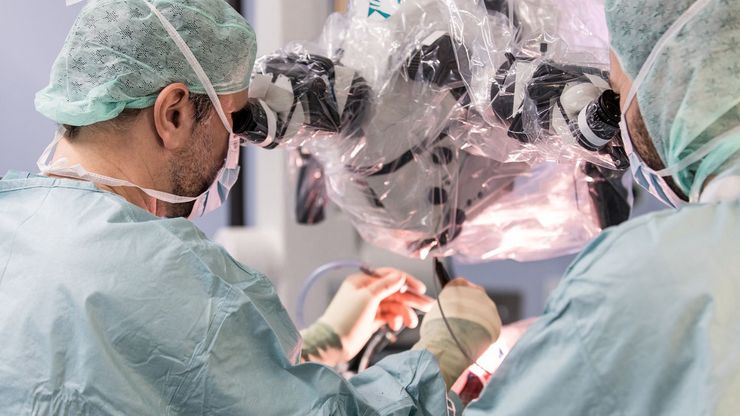
A Larger 3D Area in Focus for Neurosurgical and Ophthalmic Microscopes
Neurosurgeons and ophthalmologists deal with delicate structures, deep or narrow cavities and tiny structures with vitally important functions. Seeing a clear, large 3D area of the surgical field in…
Loading...
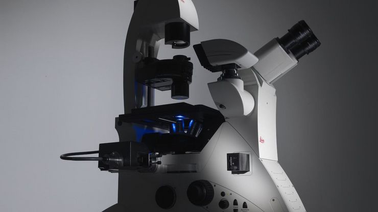
Factors to Consider When Selecting a Research Microscope
An optical microscope is often one of the central devices in a life-science research lab. It can be used for various applications which shed light on many scientific questions. Thereby the…
Loading...
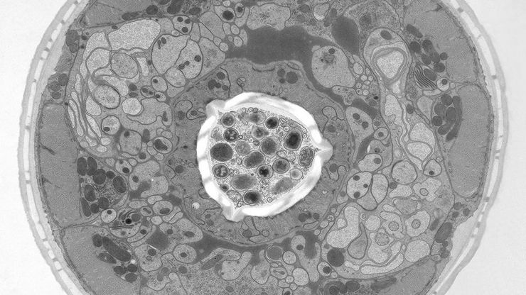
Brief Introduction to High-Pressure Freezing for Cryo-Fixation
Preparation of biological specimens for electron microscopy (EM) often requires cryo-fixation which does not introduce significant structural alterations of cellular constituents. A common method used…
Loading...

Advanced Visualization in Head and Neck Reconstructive Surgery
PRS surgeons and maxillofacial specialists face unique challenges in reconstructive procedures, particularly when operating in deep, narrow cavities and working with delicate tissues. Achieving…
Loading...
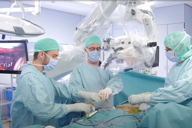
Advanced Visualization: Transforming Minimally Invasive Spine Surgery
Over the past decade, spine surgery has evolved rapidly with the adoption of minimally invasive surgery (MISS) techniques. These approaches reduce surgical trauma, speed up recovery, and help improve…
Loading...

Advanced Visualization in ENT Surgery: Prof. Darrouzet’s Expert Insights
Otologic surgery demands exceptional precision when working within the confined spaces of the middle and inner ear. Procedures such as tympanoplasty, mastoidectomy, and cochlear implant placement…
Loading...
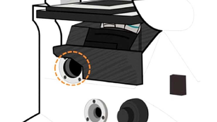
Infinity Optical Systems - From “Infinity Optics” to the Infinity Port
“Infinity Optics” is the concept of a light path with parallel rays between the objective and tube lens of a microscope [1]. Placing flat optical components into this “infinity space” which do not…
Loading...
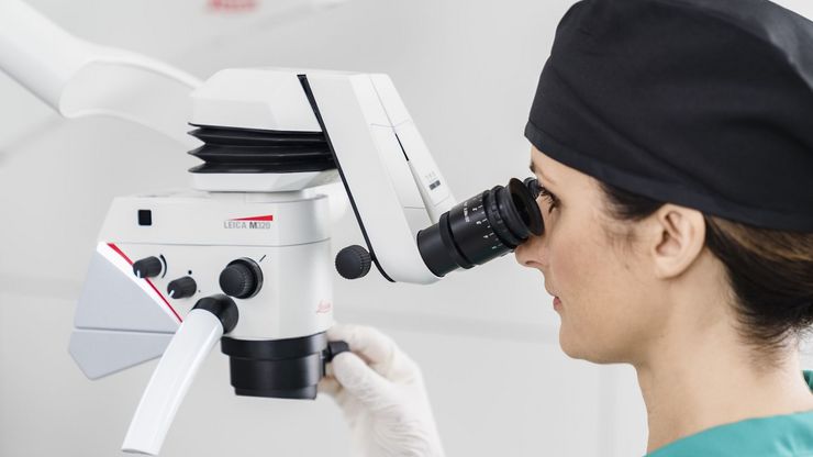
Six Features to Consider when Choosing a Dental Microscope
The dental surgical microscope has become increasingly important for high-quality and successful dental medicine, particularly in the field of endodontics. A dentist can conduct micro-invasive…
