
Science Lab
Science Lab
Bienvenido al portal de conocimiento de Leica Microsystems. Aquí encontrará investigación científica y material didáctico sobre el tema de la microscopía. El portal ayuda a principiantes, profesionales experimentados y científicos por igual en su trabajo diario y en sus experimentos. Explore tutoriales interactivos y notas de aplicación, descubra los fundamentos de la microscopía, así como las tecnologías de gama alta. Forme parte de la comunidad Science Lab y comparta sus conocimientos.
Filter articles
Tags
Story Type
Products
Loading...

Photoactivatable, Photoconvertible, and Photoswitchable Fluorescent Proteins
Fluorescent proteins (FPs) such as GFP, YFP or DsRed are powerful tools to visualize cellular components in living cells. Nevertheless, there are circumstances when classical FPs reach their limits.…
Loading...

Pinhole Effect in Confocal Microscopes
When operating a confocal microscope, or when discussing features and parameters of such a device, we inescapably mention the pinhole and its diameter. This short introductory document is meant to…
Loading...
 stereo microscope for a task like surgery.")
Rodent and Small-Animal Surgery
Learn how you can perform rodent (mouse, rat, hamster) and small-animal surgery efficiently with a microscope for developmental biology and medical research applications by reading this article.
Loading...

Navigating Through the Brain
One of the challenges of neurosurgery is orientation at the surgical site. When resecting tumors, removing arteriovenous malformations, or clipping aneurysms, surgeons often have to work near healthy…
Loading...
 illuminated with wide-band UV excitation. Note the tissue structure with blue autofluorescence. Right: Same tissue and same immunostaining with FITC label illuminated with epi-illumination using narrow")
Milestones in Incident Light Fluorescence Microscopy
Since the middle of the last century, fluorescence microscopy developed into a bio scientific tool with one of the biggest impacts on our understanding of life. Watching cells and proteins with the…
Loading...

Chronic Inflammation Under the Microscope
In the course of chronic inflammation certain body areas are recurrently inflamed. This goes along with many human diseases. With the help of widefield light microscopy, the underlying processes can…
Loading...
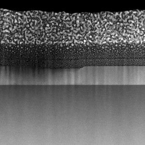
Each Atom Counts: Protect Your Samples Prior to FIB Processing
Application Note for Leica EM ACE600 - Focused ion beam (FIB) technology has become an indispensable tool for site-specific TEM sample preparation. It allows to extract electron transparent specimens…
Loading...

Factors for Selecting Student Microscopes
If chosen carefully, educational microscopes will open windows to a cosmos of minute detail which delight young minds in schools and universities – and ideally keeps them fascinated enough to make…
Loading...

Gene Editing with CRISPR/Cas9 - Breakthrough in Genome Engineering
The CRISPR/Cas9 system is one of several different bacterial systems for defense against viral attacks. It consists of two main components. One is a small piece of RNA which binds to the viral target…
