

Science Lab
Science Lab
Benvenuti nel portale delle conoscenze di Leica Microsystems. Troverete materiale didattico e di ricerca scientifica sul tema della microscopia. Il portale supporta i principianti, i professionisti esperti e gli scienziati nel loro lavoro quotidiano e negli esperimenti. Esplorate i tutorial interattivi e le note applicative, scoprite le basi della microscopia e le tecnologie di punta. Entrate a far parte della comunità di Science Lab e condividete la vostra esperienza.
Filter articles
Tag
Tipo di storia
Prodotti
Loading...

How do Cells Talk to Each Other During Neurodevelopment?
Professor Silvia Capello presents her group’s research on cellular crosstalk in neurodevelopmental disorders, using models such as cerebral organoids and assembloids.
Loading...

Overcoming Observational Challenges in Organoid 3D Cell Culture
Learn how to overcome challenges in observing organoid growth. Read this article and discover new solutions for real-time monitoring which do not disturb the 3D structure of the organoids over time.
Loading...
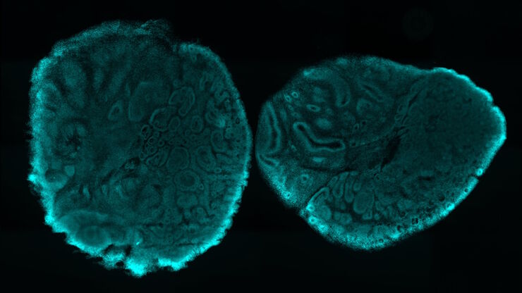
Imaging Organoid Models to Investigate Brain Health
Imaging human brain organoid models to study the phenotypes of specialized brain cells called microglia, and the potential applications of these organoid models in health and disease.
Loading...
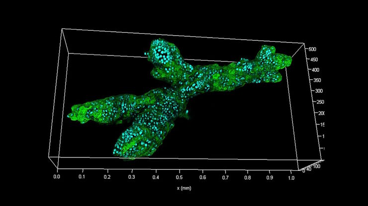
Examining Developmental Processes In Cancer Organoids
Interview: Prof. Bausch and Dr. Pastucha, Technical University of Munich, discuss using microscopy to study development of organoids, stem cells, and other relevant disease models for biomedical…
Loading...
The Role of Iron Metabolism in Cancer Progression
Iron metabolism plays a role in cancer development and progression, and modulates the immune response. Understanding how iron influences cancer and the immune system can aid the development of new…
Loading...
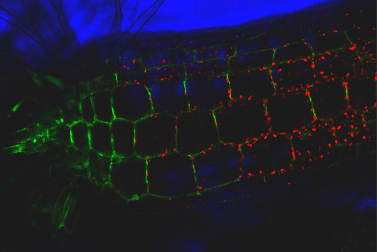
Live-Cell Imaging Techniques
The understanding of complex and/or fast cellular dynamics is an important step for exploring biological processes. Therefore, today’s life science research is increasingly focused on dynamic…
Loading...
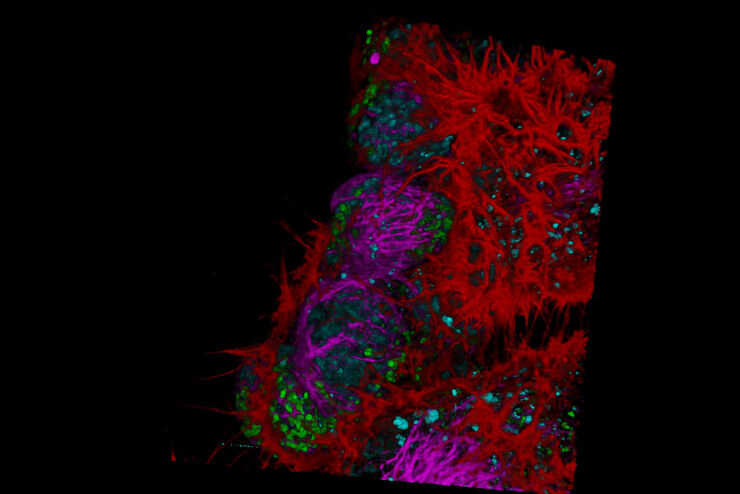
Fluorescence Lifetime-based Imaging Gallery
Confocal microscopy relies on the effective excitation of fluorescence probes and the efficient collection of photons emitted from the fluorescence process. One aspect of fluorescence is the emission…
Loading...
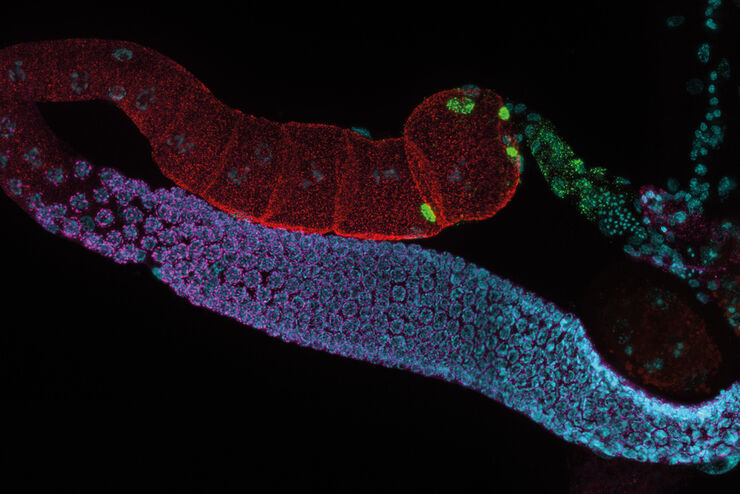
Physiology Image Gallery
Physiology is about the processes and functions within a living organism. Research in physiology focuses on the activities and functions of an organism’s organs, tissues, or cells, including the…
Loading...
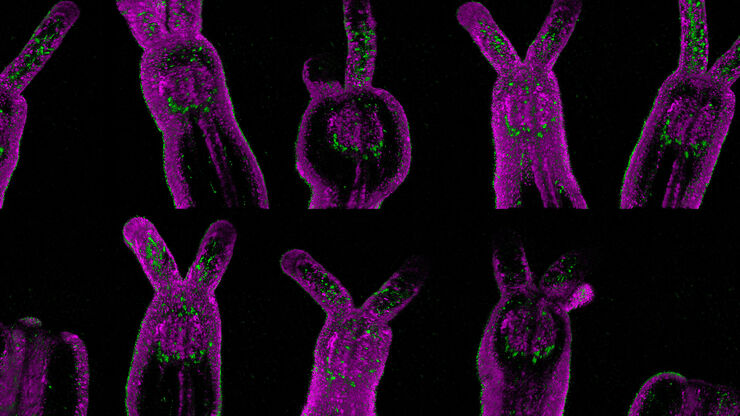
Live Cell Imaging Gallery
Live cell microscopy techniques are fundamental to get a better understanding of cellular and molecular function. Today, widefield microscopy is the most common technique used to visualize cell…


