
Science Lab
Science Lab
Das Wissensportal von Leica Microsystems bietet Ihnen Wissens- und Lehrmaterial zu den Themen der Mikroskopie. Die Inhalte sind so konzipiert, dass sie Einsteiger, erfahrene Praktiker und Wissenschaftler gleichermaßen bei ihrem alltäglichen Vorgehen und Experimenten unterstützen. Entdecken Sie interaktive Tutorials und Anwendungsberichte, erfahren Sie mehr über die Grundlagen der Mikroskopie und High-End-Technologien - werden Sie Teil der Science Lab Community und teilen Sie Ihr Wissen!
Loading...
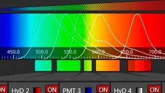
Spectral Detection – How to Define the Spectral Bands that Collect Probe-specific Emission
To specifically collect emission from multiple probes, the light is first separated spatially and then passes through a device that defines a spectral band. Classically, this is a common glass-based…
Loading...

Brief Introduction to High-Pressure Freezing
Water is the most abundant cellular constituent and therefore important for preserving cellular ultra-structure. Currently the only way to fix cellular constituents without introducing significant…
Loading...
Step by Step Guide for FRAP Experiments
Fluorescence Recovery After Photobleaching (FRAP) has been considered the most widely applied method for observing translational diffusion processes of macromolecules. The resulting information can be…
Loading...

Every Clue Counts – Forensics Inconceivable Without Microscopy
There is no crime without clues. They may be obvious, like a cartridge case at the scene of the crime or clear signs of crowbar damage on a door. But sometimes, clues are microscopically small.…
Loading...
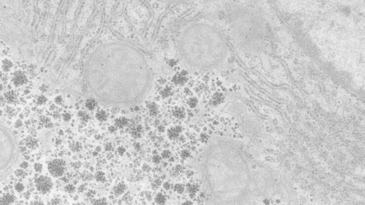
Perusing Alternatives for Automated Staining of TEM Thin Sections
Contrast in transmission electron microscopy (TEM) is mainly produced by electron scattering at the specimen: Structures that strongly scatter electrons are referred to as electron dense and appear as…
Loading...
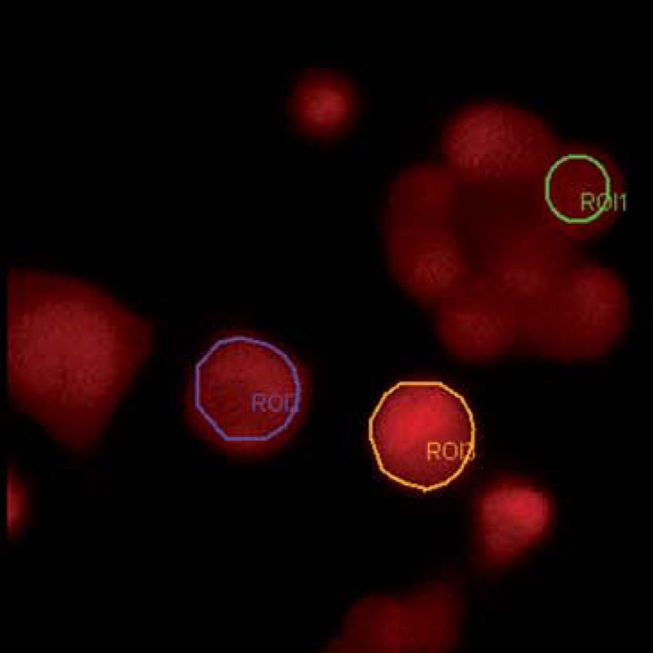
Widefield Calcium Imaging with Calcium Indicator Fura2
In eukaryotic cells Ca2+ is one of the most widespread second messengers used in signal transduction pathways. Intracellular levels of Ca2+ are usually kept low, as Ca2+ often forms insoluble…
Loading...

Brief Introduction to Critical Point Drying
One of the uses of the Scanning Electron Microscope (SEM) is in the study of surface morphology in biological applications which requires the preservation of the surface details of a specimen. Samples…
Loading...

50 Years of Image Analysis
Modern image analysis systems perform highly sophisticated image processing functions on images from an automated microscope and digital camera. 50 years ago, the first image analysis system was…
Loading...
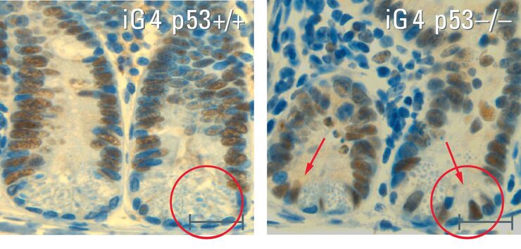
Stem Cells in the Aging Process – the Signal Protein p53 as "Guardian of the Genome"
The physiological process of aging is an elementary part of life. The processes responsible for the aging process and the effect they have is one of the key focuses of biomedical research. Dr. Yvonne…