
Science Lab
Science Lab
Das Wissensportal von Leica Microsystems bietet Ihnen Wissens- und Lehrmaterial zu den Themen der Mikroskopie. Die Inhalte sind so konzipiert, dass sie Einsteiger, erfahrene Praktiker und Wissenschaftler gleichermaßen bei ihrem alltäglichen Vorgehen und Experimenten unterstützen. Entdecken Sie interaktive Tutorials und Anwendungsberichte, erfahren Sie mehr über die Grundlagen der Mikroskopie und High-End-Technologien - werden Sie Teil der Science Lab Community und teilen Sie Ihr Wissen!
Loading...
How to Drape an Overhead Surgical Microscope
The tutorial features the Leica ARveo digital Augmented Reality microscope for complex neurosurgery. The procedure also applies to the Leica M530 OHX, OH6, OH5 and OH4.
Loading...

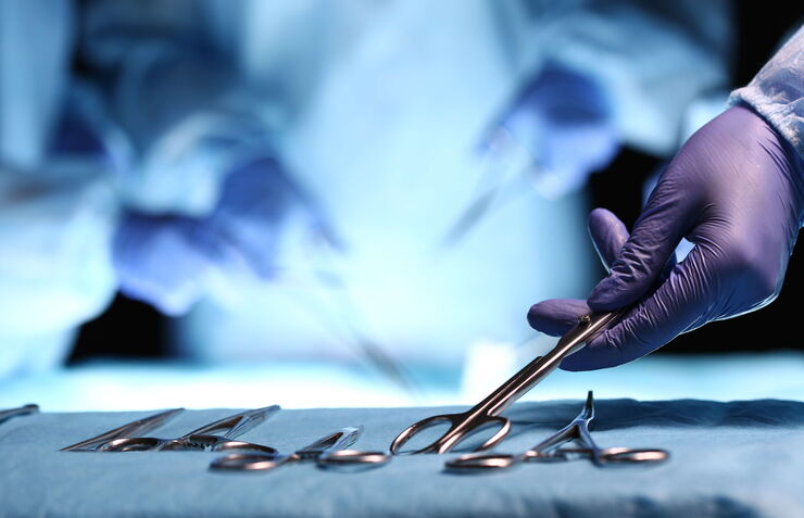
How to Drape a Surgical Microscope
Before performing surgical procedures, it is important to drape the surgical microscope to ensure sterile working conditions. At Leica, we are committed to helping you with your surgical practice. In…
Loading...
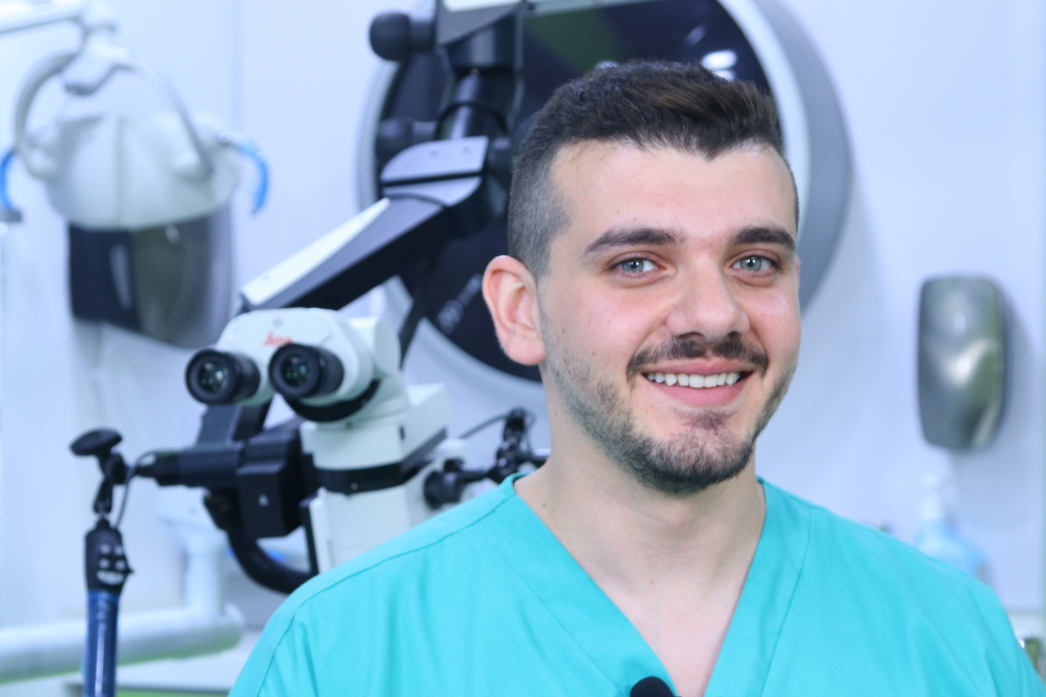
Overcoming Complexities in Microdentistry
Dr. Salam Abu Arqub, from the Smile Engineer Dental Center in Amman, Jordan, has been using Leica dental microscopes for three years for all procedures performed at the clinic. He shared his…
Loading...
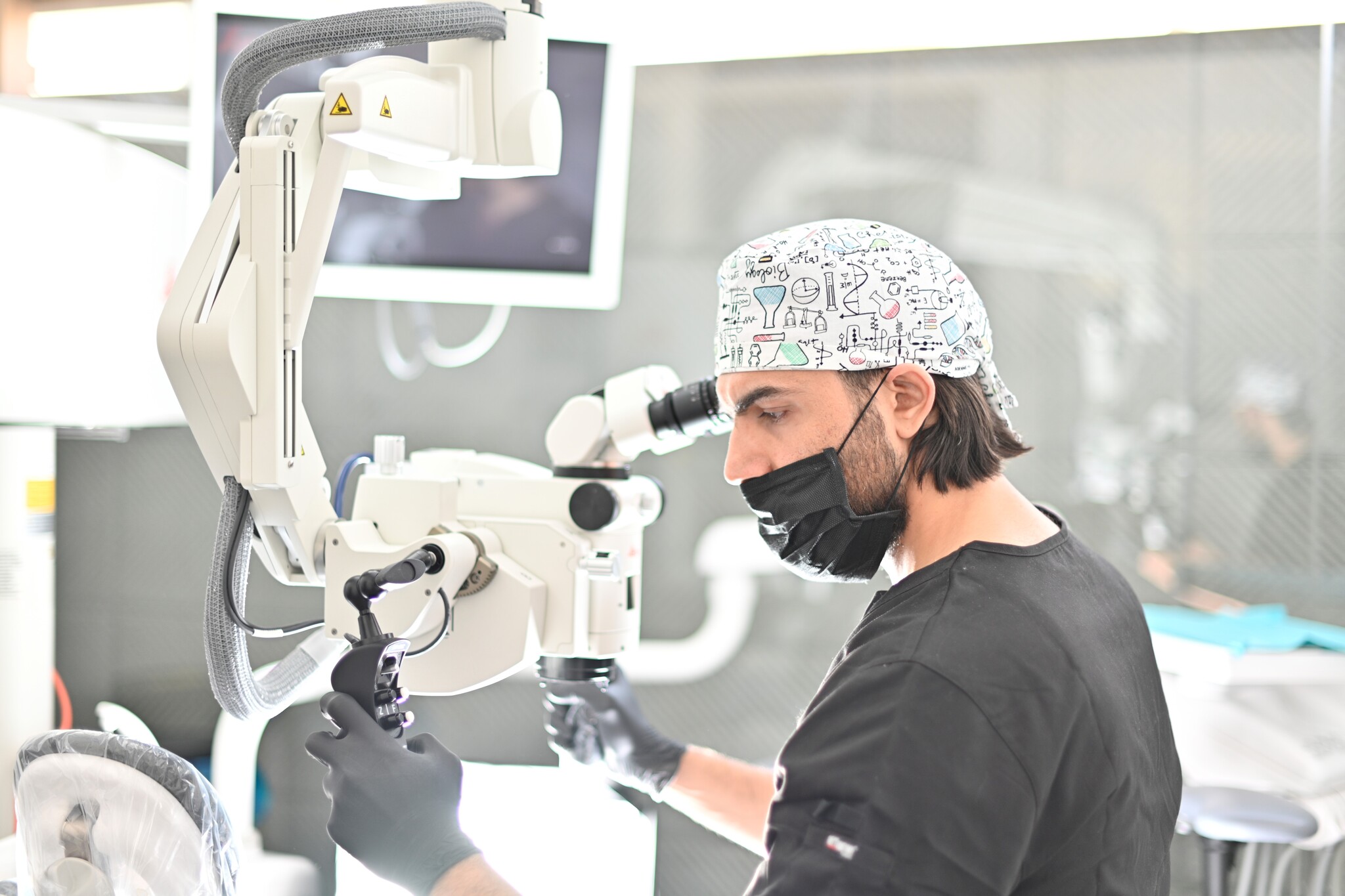
Minimally Invasive Dentistry: Visualization & Posture
Microscopes do not only provide better visualization during dental surgery. They also help ensure correct posture to avoid back pain and neck injuries. Dr. Iyad Ghoneim, from the Safad Dental Center…
Loading...
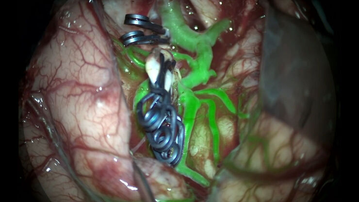
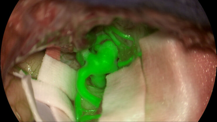
GLOW800 Augmented Reality Fluorescence in Aneurysm Treatment
This case study from Prof. Dr. Feres Chaddad talks about the treatment of unruptured MCA (middle cerebral artery) and PCOM (posterior communicating artery) aneurysms with microsurgical clipping. It…
Loading...
The Future of Fluorescence in Vascular Neurosurgery
In vascular neurosurgery, surgical microscopes are used to provide a magnified and illuminated view of the surgical field. Although surgeons benefit greatly from the superb image quality and optical…
Loading...
 stereo microscope for a task like surgery.")
Rodent and Small-Animal Surgery
Learn how you can perform rodent (mouse, rat, hamster) and small-animal surgery efficiently with a microscope for developmental biology and medical research applications by reading this article.
Loading...

Navigating Through the Brain
One of the challenges of neurosurgery is orientation at the surgical site. When resecting tumors, removing arteriovenous malformations, or clipping aneurysms, surgeons often have to work near healthy…
Loading...
What is OCT?
Optical Coherence Tomography (OCT) is a non-invasive, non-contact imaging modality used to visualize and monitor changes to the morphology of biological tissue. OCT employs low-coherence…